
overshot teeth for sale
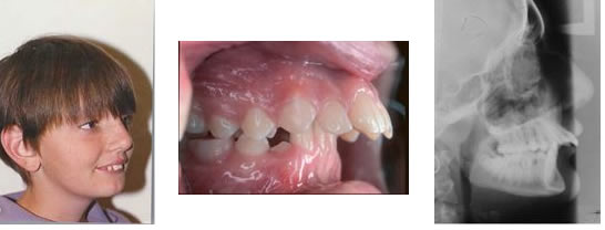
Most people aren’t born with perfectly aligned teeth. Usually, slightly misaligned teeth don’t require any medical treatment. However, correcting an underbite, especially when it’s severe, can have big benefits.
Teeth will become easier to clean. Your risks for tooth decay and gum disease will decrease. You’ll also feel less strain on your teeth, jaws, and facial muscles.
Brushing and flossing your teeth regularly in addition to visiting a dentist for checkups and cleanings are important parts of treatment for healthy teeth. But those with an underbite or other dental issues must take special care of their teeth to prevent further damage and decay.
Brush your teeth at least twice a day for two minutes each time with toothpaste containing fluoride. Pay attention to brushing along your gumline and on the inside, outside, and the back of your mouth. Be sure you floss in addition to brushing. See your dentist at least twice a year for checkups and cleanings.
Medical treatment is the only way to truly correct an underbite and align teeth correctly. At the very least, medical treatment can improve the appearance of an underbite.
In less severe cases of underbite, a dentist may be able to use wire or plastic braces or other dental appliances to move the teeth into their correct place.
Removal of one or more teeth on the lower jaw may also help improve the appearance of an underbite if overcrowding of the teeth is contributing to the issue. A dentist may also use a grinding device to shave down or smooth teeth that are large or stick out.
The earlier an underbite is addressed, the better. If a child’s underbite is less severe, parents should wait until at least age 7 to seek corrective treatment such as braces. That’s when permanent teeth begin to erupt.

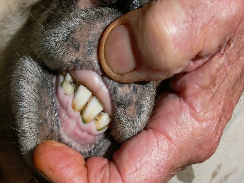
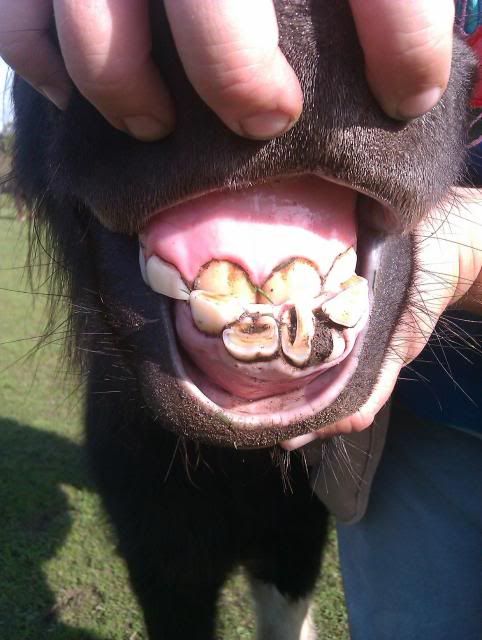
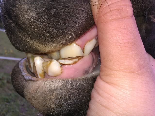
Normally, a puppy will have 28 baby teeth once it is six months old. By the time it reaches adulthood, most dog breeds will have 42 teeth. A misalignment of a dog"s teeth, or malocclusion, occurs when their bite does not fit accordingly. This may begin as the puppy"s baby teeth come in and usually worsens as their adult teeth follow.
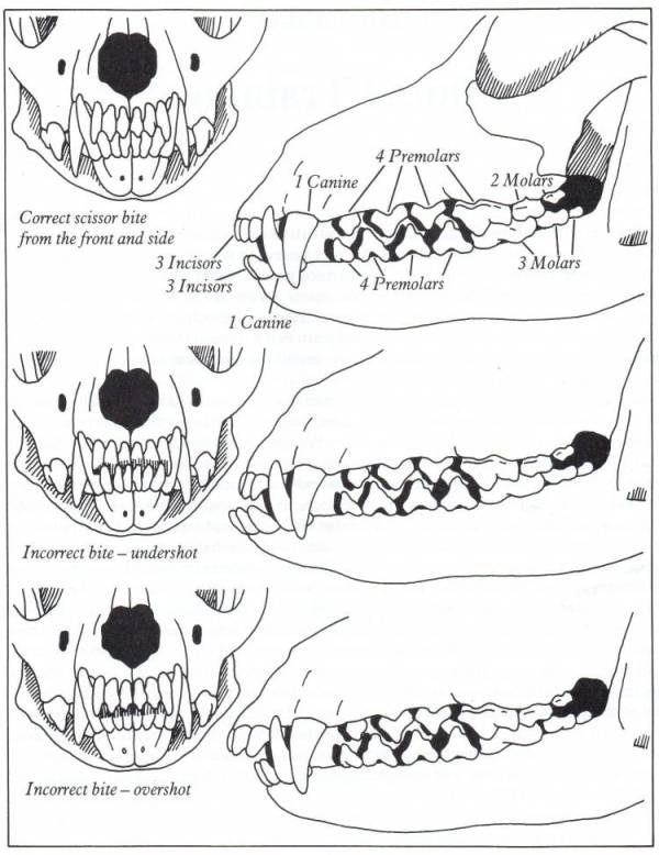
The smaller front teeth between the canines on the upper and lower jaws are called incisors. These are used to grasp food and to keep the tongue inside the mouth. Canines (also known as cuspids or fangs) are found behind the front teeth, which are also used to grasp. Behind the canines are the premolars (or bicuspids) and their function is to shear or cut food. Molars are the last teeth found at the back of the mouth and they are used for chewing.
If problems with the palate persist, a fistula may result and become infected. In cases of misaligned teeth (or malocclusion), the dog may have difficulty chewing, picking up food, and may be inclined to eat only larger pieces. They are also prone to tartar and plaque build-up.
The tips of the premolars (the teeth right behind the canines) should touch the spaces between the upper premolars, which is called the scissor bite. However, it is normal for flat-faced breeds (brachycephalic) such as Boxers, Shih Tzus, and Lhasa Apsos not to have scissor bites.
With an overbite, the upper jaw is longer than the lower one. When the mouth is closed, a gap between the upper and lower incisors occurs. Puppies born with an overbite will sometimes have the problem correct itself if the gap is not too large. However, a dog"s bite will usually set at ten months old. At this time improvement will not happen on its own. Your pet"s overbite may worsen as the permanent teeth come in because they are larger and can damage the soft parts of the mouth. Teeth extractions are sometimes necessary.
The way the upper teeth align with the lower teeth is called occlusion. It is normal for most breeds to have a slight overlap of the upper front teeth. When the jaw is closed, the lower canine (fang) should fit in front of the upper canine. Most cases of malocclusion have a hereditary link.
Most bite malocclusions do not require treatment. In some cases, extractions may be necessary. It’s a good idea to brush the teeth regularly to prevent abnormal build-up of tartar and plaque. Your veterinarian will sometimes recommend a dental specialist if you want to correct the teeth misalignment. In recent years, “braces” have been made for puppies to realign the teeth.
They are used for grasping food and help keep the tongue within the mouth. Canines (also called cuspids or fang teeth) are located on the sides of the incisors and used to grasp food. Premolars (bicuspids) are for shearing or cutting food and are located behind the canines. The molars are the last teeth in the mouth. They are used for grinding nourishment for entry into the esophagus.
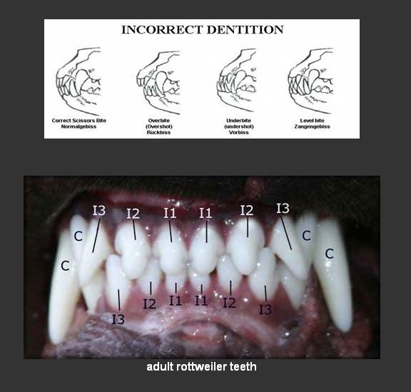
The way teeth align with each other is termed occlusion. Normal occlusion in most medium and long-nosed breeds consists of the upper (maxillary) incisors just overlapping the lower (mandibular) incisors (scissor bite). (See Photo 1.) The lower canine should be located equidistant between the last (lateral) incisor and the upper canine tooth (Photo 2). The premolar tips of the lower jaw should point between the spaces of the upper jaw teeth (Photos 3 and 4).
Malocclusion refers to abnormal tooth alignment. Overbite (overshot, class two, overjet, mandibular brachygnathism) occurs when the lower jaw is shorter than the upper (Photo 6). There is a gap between the upper and lower incisors when the mouth is closed. The upper premolars are displaced at least 25 percent toward the front when compared to the lower premolars. An underbite (undershot, reverse scissor bite, prognathism, class 3) occurs when the lower teeth protrude in front of the upper jaw teeth (Photos 7, 8) If the upper and lower incisor teeth meet each other edge to edge, the occlusion is an even or level bite (Photo 11). When the upper and lower incisors do not overlap or even meet each other when the mouth is closed, the pet has an open bite . Anterior crossbite occurs when the canine and premolar teeth on both sides of the mouth occlude normally, but one or more of the lower incisors are positioned in front of the upper incisors (Photo 12). Anterior crossbite is the most common malocclusion, is not considered genetic or hereditary and is correctable. Posterior crossbite occurs when one or more of the premolar lower jaw teeth overlap the upper jaw teeth. This is a rare condition that occurs in the larger-nosed dog breeds. A wry mouth or wrybite occurs when one side of the jaw grows longer than the other. Lingually displaced mandibular (base narrow)canines occur when the mandibular canine teeth protrude inward causing penetrating damage to the upper palate. This condition is due to either persistent primary teeth or a too-narrow mandible and can usually be corrected through an orthodontic appliance (inclined plan) used to direct the teeth into normal occlusion. Rostrally deviated canine teeth occur when the maxillary or mandibular canine is directed forward and can usually be corrected by orthodontic movement, crown reduction or extraction.
This condition is most often spotted at either the first or second puppy checks or between 6 and 8 months of age as the permanent (adult) teeth erupt. Either the deciduous or permanent lower canines occlude into the soft tissues of the roof or the mouth causing severe discomfort and, possibly, oral nasal fistulae.
The fact sheet answers many questions you may have about the cause of this problem and the various treatments available. It is important not to delay treatment of deciduous lower canines as the window of opportunity is only a matter of a few weeks until the permanent canines erupt at 22 to 26 weeks of age. A new problem can then present with bigger teeth causing more damage.
We advise you email us images of the teeth (mouth closed, lips up and side on for both left and right) just a few days before you travel. Things change quickly in growing dogs and it might save you a wasted journey.
Firstly, and most importantly, these teeth are sharp and hitting the soft tissues of the palate. These pups cannot close their mouth without pain and often hold the mouth slightly open to avoid contact. This is not pleasant. See above for an example of the damage caused to the hard palate by this problem.
Thirdly, the permanent lower canine is located lingual to the deciduous canine. This means that if the deciduous lower canines are in a poor position it is a certainty the permanent teeth will be worse. See the radiograph below. The deciduous canines are on the outside of the jaws and the developing permanent canines are seen in the jaw as small "hats". It is clear that the eruption path of the permanent canines will be directly dorsal and not buccally inclined as is normal.
For these three reasons it is advisable to surgically remove the lower canine teeth as soon as possible to allow maximum time between the surgery and the time the permanent teeth erupt at between 22 and 24 weeks of age. See our file for illustration of removal of deciduous canines.
The permanent successor tooth is located lingual to the deciduous tooth and wholly within the jaw at this stage. Any use of luxators or elevators on the lingual half of the deciduous tooth will cause permanent damage to the developing enamel of the permanent tooth. See the images below showing canines (and also the third incisor) with extensive damage to the enamel. The radiograph also shows how much damage can occur to the teeth - see the top canine and adjacent incisor. Some severely damaged teeth need to be extracted while other can be repaired with a bonded composite. This damage is avoidable with careful technique using an open surgical approach.
Surgery to remove the deciduous canines may not prevent to need for surgery on the permanent canines but, without it, few cases will resolve if left to nature. Many owners are reluctant to have young pups undergo surgery. Our view is that surgical removal of the lower deciduous canines will not guarantee the problem does not happen again when the permanent teeth erupt but without surgery the chances are very slim.
If the permanent teeth are lingually displaced the pup is usually older than 24 weeks. The trauma caused by the teeth on the soft tissues can be considerable with pain as a consequence.
Do not try ball therapy with deciduous (puppy) teeth. There are two main reasons for this. Puppy teeth are fragile and can easily break. More importantly, the adult canine tooth bud is developing in the jaw medial to the deciduous canine tooth (see radiograph above in the puppy section). If the deciduous crown tips outwards the root will tip inwards. This will push the permanent tooth bud further medial than it already is.
Ball therapy will only work with adult teeth and only in some cases where the lower canines have a clear path to be tipped sideways - laterally - through the space between the upper third incisor and canine. The window of opportunity can be quite short, around 6 weeks, and starts when the lower canine teeth are almost making contact with the hard palate.
The size and type of the ball or Kong is critical. The ball diameter should be the distance between the tips of the two lower canine teeth plus 50%. Therefore if this distance is 30mm the ball diameter is 45mm. If the ball is too small it will sit between the lower canines and produce no tipping force when the pup bites down. Too large a ball can intrude the lower canines back into their sockets.
These permanent teeth can theoretically be treated by three options. Not all options are available to all cases. These options are described below and are either surgical removal of the lower canines teeth (and possibly incisors also), crown amputation and partial pulpectomy or orthodontics via an inclined bite plane bonded to the upper canines and incisors. The latter option may not be available to all dogs if the diastema (space) between the upper third incisor and canine is too small for the lower canines to move into or if the lower canines are located behind (palatal) to the upper canines.
Radiograph left lower canine before (left) and immediately after (right) surgery. Note the immature morphology of the canine teeth - thin walls and open root apices.
In order to monitor this process of maturation we need to radiograph these teeth twice at 4-6 months post-op and again at 12 -16 weeks post-op. This is a mandatory check. The quoted success reate of 92% implies 8% failure. Half of those to fail in the Luotonen study happened over a year post-op. To ensure any failure of maturity is identified we will not perform this surgery unless the owner agrees to this.
Normally a composite resin bite plane is bonded onto the upper teeth (see below) with an incline cut into the sides. The lower canine makes contact with the incline when the mouth closes and, over time, the force tips the tooth buccally. This takes around four to eight weeks. The lower canine will often migrate back into a lingually displaced position when the bite plane is removed. This can occur if the tooth height of the lower canine is too short (stunted). If the lower canine is not self-retained by the upper jaw when the mouth is shut further surgery may be required.
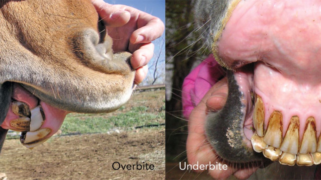
An overshot jaw is when the upper jaw extends further out than the lower jaw, with the horse having an overbite (also called a parrot mouth). When the lower jaw extends farther out than the upper jaw, we call this an undershot jaw,with the horse having an underbite (also referred to as a monkey jaw or sow mouth).
As far as genetics go, parrot mouth is NOT directly inheritable. It is rare to see an individual sire or mare throwing an abnormally high number of parrot mouthed foals. The most common cause of it is when a mare is bred to a stallion with a very different head type than she has. Surprisingly, these two animals often have normal teeth structure, yet when they are bred, the mismatch is so great that a parrot mouthed offspring is produced.
It is important to remember that malocclusions in horse’s teeth (when they are not in the correct positioning and alignment) is poorly understood when it comes to how inheritable it is, and is often a very complex mismatch of many genes. Having a badly malformed mouth in your horse may present a risk in breeding, but certainly doesn’t mean that animal will throw offspring with a similar condition.
Horses’ teeth do not grow indefinitely like rabbits’ teeth, but for some time they continue to erupt into the mouth — with the purpose being to replace the tooth which is worn away during the chewing process.
The eruption process of teeth works fine if the teeth all line up even and oppose one another. But if they are not matched, then the tooth which is not opposed will continue to erupt into the mouth and become longer and longer.
As the elongating tooth or teeth become more prominent, they may cause the tooth to be moved or forced out of its normal position and may also restrict the whole jaw’s normal RCM (rostro-caudal movement) while eating or when ridden.
In the past three articles we have discussed the importance of canine posture, how it influences health and soundness, and what are the most important factors controlling posture: the neck, the feet and the teeth.We learned that manual therapy can help reset the proprioceptors of the neck after injury, and that keeping toenails short can have a profound effect on back and hind end pain and disability.What about the teeth?And what could we possibly do about them that would be ethical in the show ring?
Skull shape is one of the most biologically important variations in the dog, because changing the “default” cone-shaped head will change the size and shape of the brain case, the eyes, nose, teeth and airway.There are some health risks that are suspected to have associations with the size and shape of the dog’s head. Researchers are currently trying to understand the causes of Syringomyelia (SM), a common spinal cord abnormality in small breed dogs. It is believed that genetic factors contribute to the disease.
In a very broad sense, we have three basic skull types in domestic breeds: long nosed (dolichocephalic), short-nosed (brachiocephalic) and medium (mesocephalic).The dolichocephalic breeds, like Greyhounds and Borzois, tend to have very narrow skulls, and may have problems with eye formation, overbites and not enough room for their incisor teeth to fit properly.Brachiocephalic breeds, like Pugs and Bulldogs, have underbites, which are even more exaggerated in some versions of these breeds.When the shape of the skull is distorted, the space into which the teeth erupt can be distorted as well.This results in crooked teeth, that don’t fit together properly, or “malocclusions.”
Why do dog breeders care about bite?Because well-bred, truly functional dogs have good bites! A good bite is associated with good posture and good gaiting, because the teeth and temporomandibular joints (TMJ) are giving critical postural information to the brain.A good bite results in neutral TMJs, which allow neutral posture.Try this exercise: Stand on level ground with easy neutral stance, arms at your sides.Feel how your weight is centered between your feet.Thrust your lower jaw forward as far as you can voluntarily, creating an underbite.Wait, and feel the postural changes.Now pull the jaw back as far as you can.Most people will feel their bodies pitch forward and back with the movement of the jaw.You can experiment with side to side as well, and feel your weight shift from foot to foot.This is a cool “party trick,” but it actually shows something very profound: jaw position helps determine weight-bearing, because the top priority of the nervous system is to keep the brain safe by making sure the nearby TM joints are symmetrically stimulated, indicating that the head is level and symmetrically supported.When a dog has a congenital or genetic malocclusion, the rest of the body may have an adapted posture-- which may make them susceptible to some weight-bearing injuries over time.
What about dental anomalies outside the brachiocephalic/dolichocephalic pattern?While orthodontic procedures can help some adult dogs become more functional, it is considered unethical to use these techniques on a potential breeding animal.But some dental problems are from juvenile injury, and can be helped with early intervention.It is critically important to evaluate the baby teeth at six weeks, because missing teeth and non-symmetrical jaw growth can be most easily influenced in the fast growing young dog.Why should we do this?Cutting edge research in epigenetics shows that life experience influences gene expression in a heritable way. And it will improve a dog’s quality of life, and athletic performance to have a functional bite.A truly functional bite is self-cleaning, requiring less dental intervention.And it will help reduce the risk of musculoskeletal problems secondary to postural abnormalities, like hip dysfunction, ACL tears, arthritis, and disc disease.
Here, Paddy Dixon, MVB, PhD, MRCVS, professor of equine surgery in the University of Edinburgh’s Division of Veterinary Clinical Sciences, describes common problems horses encounter as their teeth develop, what problems these abnormalities cause, and treatment options.
“Many horses have some degree of overjet,” Dixon says. An overjet, also referred to as an overshot jaw, occurs when the chewing surfaces of the upper incisors (the front teeth) lie slightly in front of the chewing surfaces of the lower incisors. Most times, this slight misalignment does not cause ill effects to the horse.
“If very marked and left untreated in foals, cases of severe overjet may develop into overbite,” he cautions. This condition, commonly referred to as “parrot mouth,” occurs when all of the upper incisors lie in front of the lower incisors, resulting in excess tooth growth on the top row of teeth; this excess tooth growth can sometimes cause the horse to develop a smilelike appearance when the central upper incisors overgrow .
“Overjet and overbite are aesthetically undesirable but surprisingly, these problems rarely cause difficulty in prehension (grasping food),” Dixon notes. If the horse develops these smilelike incisors, veterinarian should evaluate and reduce the excess tooth gradually, ensuring that the underlying pulp is never exposed or overheated during the process. Most horses affected with overjet and overbite also have their top row of cheek teeth slightly in front of the lower cheek teeth, and thus, they develop overgrowths on the front of their first upper cheek tooth and the back of their last lower cheek tooth, as discussed below.
Dixon explained that with both underjet and overjet, the main clinical concern is a misalignment of the horse’s six cheek teeth (three molars and three premolars).
It’s a mouthful to say, and rostrally positioned (positioned closer to the nose) upper cheek teeth can cause a mouth full of problems for horses. Dixon explains that an imbalance in growth rates between maxillary (cheek bone) and mandibular (lower jaw) bone cause this disorder, which is “nearly always associated with incisor overjet or overbite.”
Several problems can arise from the upper cheek teeth being positioned further ahead than the lower ones. “Because the maxillary and mandibular cheek teeth rows are not in full occlusion (bite alignment), localized cheek teeth overgrowth–colloquially termed beaks, hooks, or ramps–develop on the front of the upper cheek teeth,” Dixon explains. “These overgrowths may be pushed against the lips and cheeks by the bit or noseband and so cause mucosal ulceration and bitting problems.”
Wry nose is a condition in which an affected foal’s upper jaw and nose are deviated, or turned to one side, Dixon explains. The teeth associated with this condition always have malocclusion (poor alignment), although most foals can still nurse and in most cases are bright and active.
This condition–in which one or more of an affected horse’s teeth do not develop–has the potential to affect both deciduous (baby) and permanent teeth, although “true” hypodontia most commonly affects permanent teeth, Dixon explains. And while a missing tooth might not seem like a major problem, Dixon explained that its absence can cause long-term damage to the horse’s mouth.
“Developmental hypodontia is relatively uncommon in horses, with the absence of equine teeth usually due to traumatic loss, disease, or age-related wear,” he explains. The main problem associated with missing teeth is overgrowth of the opposing tooth. In these cases overgrowth can be managed with semiannual dental care to prevent the overgrown teeth causing damage to surrounding structures
“The presence of supernumerary (additional) teeth is relatively uncommon in horses, usually developing in the permanent dentition,” says Dixon. He added that supernumerary teeth can be comprised of a single tooth or more than one tooth joined together.
“Equine supernumerary incisors may be more common–or possibly more readily identified–than supernumerary cheek teeth,” Dixon notes. Extra incisors often cause overcrowding and displacement of the normal teeth. In some cases he suggested it is possible to remove the additional teeth using sedation and nerve blocks. “If (the supplemental teeth are) interwoven amongst the other incisors, differentiation of supernumerary and normal teeth is difficult and safe extraction may be impossible,” he notes. In these cases regular veterinary dental care can help keep the horse’s mouth as healthy as possible.
Supernumerary cheek teeth are relatively uncommon in horses and can cause overcrowding and irregular spacing between teeth leading to periodontal disease. Treatment depends on the location of the supernumerary teeth and what problems they cause, he noted. Following careful clinical examination and radiography (X rays), common treatment options include extracting the additional tooth, widening or filling abnormal interdental spaces (that develop painful food packing), and reducing overgrowth.
“Dysplasia, or abnormal development of teeth, can involve the crown, roots, or all parts of the tooth,” Dixon says. “Dysplasias in the gross anatomy include dilacerations (abnormal bending of teeth), double teeth, abnormalities of size, and concrescence (roots of adjacent teeth joined by cementum) of teeth.”
Additionally, he notes that teeth can develop either too large (macrodontia) or too small (microdontia). Amelogenesis imperfecta (a tooth development disorder in which teeth are covered with thin, abnormally formed enamel) is another rare dental disorder in horses, Dixon added.
“Some cases of ‘stepmouth’ and ‘wave mouth’ are caused by mismatched eruption times of opposing permanent cheek teeth, allowing overgrowth of the teeth which erupt first,” Dixon explains.
Smaller and miniature breeds have this type of disorder more commonly than other breeds due to a likely mismatch between the size of their teeth and of the support bones (jaws), Dixon noted, and the steplike or wavelike overgrowths can remain–or even increase in size–throughout an affected horse’s lifetime.
Dixon notes that both deciduous incisors and cheek teeth can be retained past their normal time of shedding. In some cases, he noted, this will cause the permanent tooth to emerge into a displaced position adjacent to the retained deciduous tooth. The newly erupted tooth is often not fully aligned with its opposite tooth and so develops an overgrowth that can even cause chewing restrictions.
In most cases, Dixon says, these retained deciduous teeth can be readily identified and removed. However, it is sometimes advisable to have a veterinarian take a radiograph prior to extraction to ensure the additional tooth is not of the supernumerary variety.
Light breeds and miniature horses are also prone to developing cheek teeth vertical impactions (teeth that are too tightly squeezed by their neighbors and cannot freely erupt into the mouth) when permanent teeth are emerging. However, many larger breeds such as Thoroughbreds also develop this problem. They most commonly cause painless swelling on the bottom of the lower jaw (i.e., eruption cysts, or “3-year-old bumps” and “4-year-old bumps”) that usually disappear after a year or so, because “as the jaw bones lengthen, the impacted cheek teeth have room to erupt normally,” Dixon says.
If there is not enough room for the fourth cheek tooth to erupt normally at one year of age or for the fifth cheek tooth to erupt at two years of age, the (overcrowded) erupting tooth might be displaced and cause a potentially lifelong problem. The displaced tooth might then grow toward the cheeks or tongue, causing painful inflammation, but the most significant problem will be the presence of diastemata (space) between the normal teeth row and the displaced tooth. At these sites, food invariably becomes impacted and the horse can develop painful periodontal disease. If the displacement is very severe, a veterinarian might extract the displaced teeth–a procedure most readily performed in older horses, especially when deep periodontal disease is present.

Persian cats and some other breeds are brachycephalic; "Brachy" means "shortened" and "cephalic" means "head". The skull bones of brachycephalic cats are shortened in length, giving the face and nose a "pushed in" or "flat" appearance. Due to the shorter bones of the face and nose, the anatomy and relationships with other soft tissue structures are altered. These breeds can be subject to teeth and jaw problems that can include crooked teeth. Sometimes baby teeth can be crooked but the adult teeth grow in normally.
The entire area of the mouth is referred to as the "jaw" and consists of the Maxilla (the upper jaw) and the Mandible (the lower jaw). When the upper jaw protrudes it is referred to as "overshot" (dental term) or an "overbite" (layman"s term). If the lower jaw protrudes it is called either "undershot" or an "under bite".
In animals with an overshot or undershot jaw this condition is also known as "Asymmetrical Jaw", meaning the jaws are not identical (one is longer than the other). While not as common in the Persian breed, a twisted mandible can happen whereby the two jaws do not touch each other and the teeth may protrude outside of the lips. Sometimes causing the teeth to penetrate the roof of the mouth, jab, or poke the gums in different places.
Improper alignment of the jaw can cause soft tissue trauma from the canine teeth (the four long, sharp, fang-like teeth). If a jaw is severely undershot, it may cause trauma to the lip as well. Poorly aligned teeth can have serious periodontal implications which can include:
Soft tissue trauma around the canines. The animal"s gums can be red and/or inflamed, and sometimes lesions in the soft tissue area can result due to friction and pressure of the teeth. In extreme cases, the canine teeth can also erode the palate and cause food to enter the nasal cavity.
In an extreme case where the bite and/or jaw is so severely misaligned that it interferes with eating and/or chewing, there are treatments once the adult teeth are in. These include:
A dental veterinarian (this is a specialist, not a regular veterinarian) can apply rubber bands to the cat"s teeth to bring them back to normal alignment. Retainers and other devices can also be used. Personally, I would never go to this expense.
PLEASE NOTE: The treatments described above are for "EXTREME" cases where eating or chewing are impaired due to the misalignment of the jaws or crooked canine teeth. More often than not, an Asymmetrical Jaw does not require any outside intervention.
It is very important that owners pay close attention to a young kitten"s teeth and the alignment of their jaws prior to their adult teeth coming in. Development of a potentially incorrect bite (Asymmetrical Jaw) may be corrected by the simple removal or clipping of the upper or lower canines (depending on whether it is undershot or overshot). Retained baby teeth can also cause misalignment of the jaw or crooked canine teeth as the adult teeth come in.
Teeth strongly developed, powerful canine teeth fitting closely. Jaws strong, with a perfect, regular and complete scissor bite, i.e. upper teeth closely overlapping lower teeth and set square to the jaws. Complete dentition important.
The correct mouth should be a “Scissor” bite, with closely overlapping top and bottom canine teeth. Any deviation from this is a fault; for example an “overshot” jaw where the top teeth overlap the bottom teeth by a significant margin, or an “undershot” jaw, where the top teeth close behind the lower incisors. An overshot jaw is more common than an undershot jaw. Neither of these faults is likely to be a problem for a Dachshund living as a pet, but he would not be able to grasp and hold his prey firmly if he was a working dog with either of these faults.
The extent of the scissor bite is sometimes a cause for discussion. Judges who own Wires have been known to observe in their critiques that the teeth of the other varieties are overshot and, conversely, judges who own Smooths or Longs have criticised Wires for having pincer bites. If the top teeth closely overlap the bottom teeth, the dog has a correct bite. A pincer bite would have edge-to-edge teeth.
Complete dentition means there should be 22 lower teeth and 20 upper teeth. There should be 6 incisors in each jaw. Occasionally, one incisor might be missing, but sometimes two are missing; both situations are faults. There should be 2 canines in each jaw – 1 on each side. There should be 8 pre-molars in each jaw – 4 on each side; missing pre-molars would also be considered to be faulty. There should be 4 molars in the upper jaw – 2 on each side – and 6 in the lower jaw – 3 on each side. Note that missing premolars is not considered to be a fault under the latest FCI Standard.
A full set of teeth means the dog can grip strongly and if you try to prise a Dachshund’s jaws apart you should be able to feel how powerful his jaws are. This applies as much in the Miniatures as the Standards; they should not have soft, toy-ish mouths, or weak jaws.
Continental judges, used to the FCI Standard, seem to be more rigorous than UK judges when examining the teeth. Clearly, more than a cursory glance is required, but heavy-handed probing is not fair on the dog.
Luckily, breeders seem to have corrected the crowded teeth and narrow jaws that were seen in the Mini Wires not so long ago, but judges do need to be vigilant and observations on dentition in their critiques can be helpful.


 8613371530291