
flag pup joint in stock

A joint of tubing or casing included in the string at a known position to provide a reference point for further operations. A short pup joint that registers clearly in a collar locator log is a common flag joint.

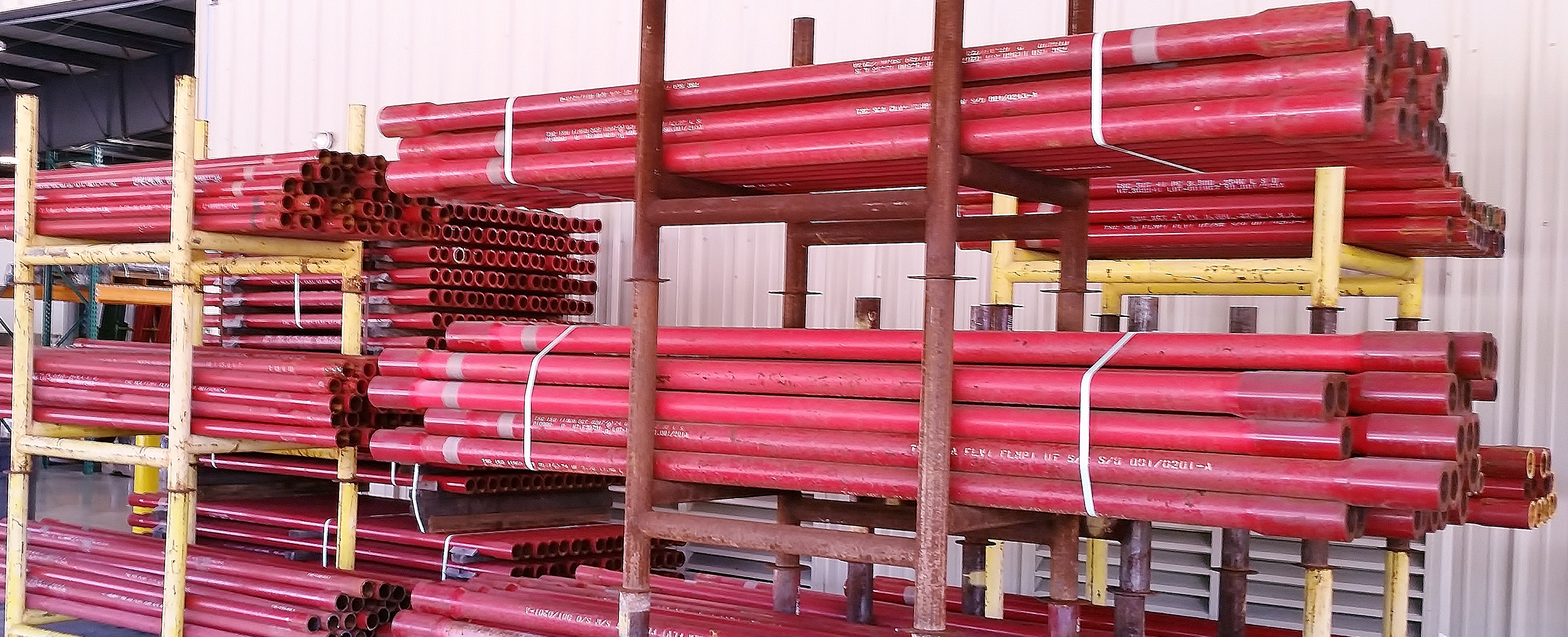
A pup joint is Casing, Pipe or Tubing shorter in length than a standard tubular string. This allows for the adjustment and installation of tools and various tubular components when placement downhole is critical for a specific project. A Spacer Pipe is another reference used to identify pup joints. Pup joint features consist of connections, lengths, weights and material grade.
Crossover pup joints are manufactured from seamless mechanical tube. As with all Crossover products, each piece is marked with a distinctive job number and heat number that is fully traceable. A complete range of sizes (1" to 4.5"), weights (standard or heavy wall), and grades (J-55, N-80, L-80, and P-110) are commonly available from stock in 2", 3", 4", 6", 10", and 12" lengths. Lengths up to 20" are available upon request.
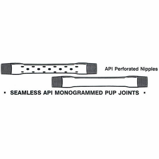
Seamless pup joints with premium connections are available in API and exotic alloy grades. Premium ends are threaded by the manufacturer or authorized licensee.
Available with standard or special perforation spacings. Each joint has four rows of ⅜ inch holes drilled longitudinally along the tube. Optional patterns, hole size, and lengths furnished upon request.
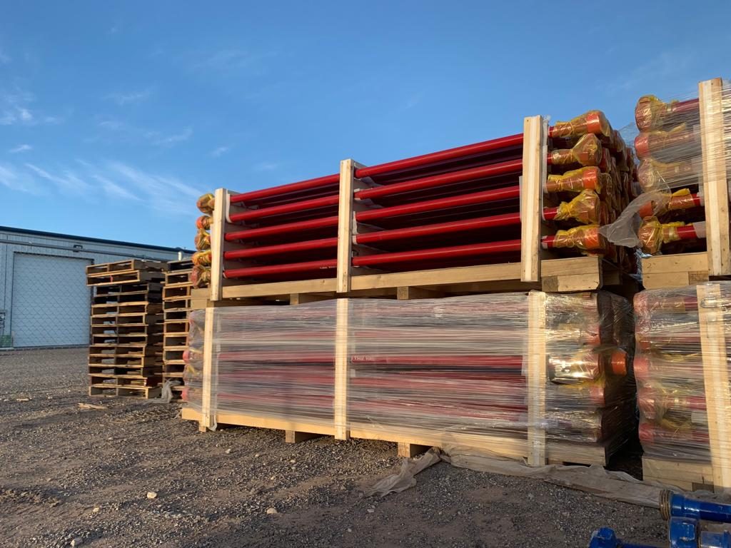
A blast joint is shorter in length than standard tubular joint. Built with a heavy wall pipe it is incorporated in the production string to facilitate production across any perforated interval and zone. Blast Joints are manufactured to the following specs connections, lengths, weights and material grade.
Crossover Blast Joints are heavy wall pin by box connectors used in tubing strings and are designed to minimize the effect of external erosive action caused by production fluids. Blast joints are located opposite the location of perforations in the production casing or just below the tubing hanger in sand frac designs. Crossover blast joints are manufactured from seamless mechanical tube in sizes ranging from 2 3/8” to 4 1/2” OD. Any length, grade of material, and threading is available at the customers request. Typical lengths are 10" and 20". Both API and Premium threads are available.
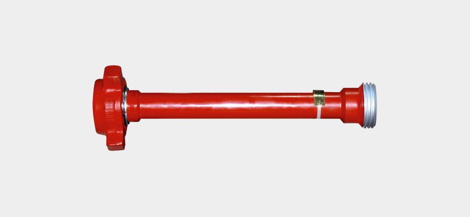
Crossover Coarse Thread Tubing Safety Joint provides for emergency recovery of the major portion of the tubing string should it become necessary to abandon the equipment below. Precision left-hand threads facilitate the release of the joint by right-hand tubing rotation. Equipment requiring right-hand rotation should not be used below the Safety joint.
Crossover Straight-Pull/Shear-Out Safety Joint is used between packers in dual and triple completions and in selective completions using Hydrostatic Single-String Packers. It is also used when rotational releasing is not desired. When ran above the upper packer in a single-string completion, however, the shear value should be adjusted to compensate for any hydraulic conditions that exist when the string is landed, or that are created by well treating operations. They are available in keyed and non-keyed configurations.
Crossover rotary shoes are manufactured from specially tempered steel to provide the ultimate in toughness and durability. They are used to cut a clearance between the fish and the wall of the well bore. Each shoe is tailored to fit a particular downhole need and normally is run on the bottom of one or more joints of washover pipe. Shoe design is dictated by whether it cuts on the bottom, on the OD, on the ID, or any combination of these. When hole sizes permit, additional clearances can be cut using side ribs, thus providing greater circulation.
VetriScience® Laboratories GlycoFlex® Classic 600 mg is a chewable tablet that supports joint and connective tissue function in canines. It is the foundation for our entire GlycoFlex® Stages line of hip and joint support for pets and, is recommended for joint health maintenance programs for puppies and dogs throughout their lives.
GlycoFlex® hip and joint support for dogs contains perna canaliculus, an edible green-lipped mussel sourced from the waters of New Zealand. The perna has been freeze-dried so that the whole organism is included, providing many of the compounds necessary to maintain healthy connective tissue. It is rich in amino acids, polypeptides, naturally chelated minerals, fatty acids, glycosaminoglycans (GAGs), vitamins, glycoproteins, protein complexes, polysaccharides and the nucleic acids RNA and DNA. Perna contains all major classes of GAGs including chondroitin 4 and 6 sulfates and hyaluronic acid that occur in connective tissue such as joint cartilage, tendons, ligaments, and synovial fluids. The compounds provided from Perna also are known to support other systems in the body including sperm motility, vascular system, bladder function, immune system function, and gut health.
Hip dysplasia is abnormal development of the coxofemoral joints. Hip dysplasia occurs principally in large dogs but also affects small dogs and cats. The condition is typically bilateral but can be asymmetric.
Hip dysplasia is an inherited disorder. Heritability estimates range from 0.2 to 0.6. With the use of more sensitive radiographic interpretation and newer methods of imaging the coxofemoral joints, estimated heritability in German shepherd dogs has been raised from 0.46% to 61%.25 Environmental factors influence the phenotypic expression of hip dysplasia. Overnutrition is one of the principal nongenetic factors that influences expression of canine hip dysplasia.26 Hip dysplasia is a developmental, age-related disorder; and the phenotypic manifestations are not present at birth. A variable amount of time must elapse before radiographic changes manifest. Once present, these radiographic changes usually progress with age.
The earliest recognizable changes in the coxofemoral joints are a combination of perifoveal cartilage erosion, hypertrophy of the round ligament of the femoral head, synovial effusion, and synovitis.27 These changes cannot be identified radiographically, but the strongest clue to their presence can be obtained by testing for signs of joint laxity, which appears to be precipitated by synovial effusion. Joint laxity may be palpated and visualized radiographically (Figs. 21.26
) developing as (1) perichondral osteophyte formation, (2) remodeling of the femoral head and neck, (3) remodeling of the acetabulum, and (4) increased opacity of subchondral bone of the femoral head and acetabulum. A line of enthesophytes on the caudal aspect of the femoral neck, termed the Morgan line, has been described as an early sign of coxofemoral degenerative joint disease (Fig. 21.29
).30 As the degenerative process advances, the femoral neck becomes thickened, and the surface of the neck becomes irregular as a result of the growth of a collar of perichondral osteophytes. The acetabulum loses its cuplike shape and becomes shallow. Increased bone opacity of subchondral articular surfaces represents bone sclerosis, which is a response to cartilage thinning. Subchondral cyst formation is an infrequent manifestation of degenerative joint disease in small animals but may be observed occasionally.
Normal mature coxofemoral joint. Note that at least half the femoral head lies medial to the dorsal acetabular margin (white arrows). The cranial margin of the femoral head is separated from the adjacent acetabulum by a fine radiolucent line, which represents the joint cartilage and a microfilm of synovial fluid (black arrows). The shape of the radiolucent joint space is symmetric.
Moderate hip dysplasia. Subluxation of the femoral head is accompanied by remodeling of the acetabulum. The cranial acetabular margin is angulated (black arrow), and the acetabulum is shallow. Note the wedge-shaped joint space (white arrows) created by subluxation of the femoral head.
Moderate coxofemoral degenerative joint disease. The acetabulum and the femoral head have undergone advanced remodeling. Osteophytes have formed on the femoral neck and head, as well as on the cranial acetabular margin. New bone formation has filled the acetabular fossa, and the opacity of acetabular subchondral bone is increased.
An early sign of degenerative joint disease is the Morgan line, representing enthesophyte formation on the caudal aspect of the femoral neck, medial to the trochanteric fossa (black arrow).
Feline hip dysplasia. A, “Normal” is compared with varying degrees of coxofemoral subluxation (B and C). D, Degenerative changes, with osteophyte formation on the cranial effective acetabular margin (white arrow), are a typical manifestation of feline coxofemoral degenerative joint disease.
Methods of evaluating the coxofemoral joints can be divided into those that identify joint laxity proactively and those that examine for radiographic evidence of degenerative joint disease. Phenotypic screening programs used internationally fall into the latter group and rely on assessment of the extended ventrodorsal radiographic projection (Fig. 21.32A
), although this projection is an insensitive indicator of coxofemoral joint laxity. To identify coxofemoral joint laxity reliably, a stressed ventrodorsal projection is used. With the femurs in a distracted position (see Fig. 21.32B), coxofemoral laxity can be quantified and the calculated laxity index (i.e., Distraction Index) used to rank individual dogs within their breed with respect to hip joint tightness or looseness.32 The Distraction Index (DI) is also a useful indicator of the likelihood of future coxofemoral degenerative changes. This information can be obtained at a much earlier age by using distraction radiography than with the standard extended ventrodorsal projection. Assessing coxofemoral laxity is an important component of complete radiographic assessment of the coxofemoral joints.
A, Extended ventrodorsal projection of the coxofemoral joints (Orthopedic Foundation for Animals [OFA] preferred view). Note bilateral symmetry of the pelvis and femurs. The coxofemoral joints appear normal. B, Ventrodorsal distraction projection (PennHIP view) of the same dog. Bilateral coxofemoral laxity is evident.
Many screening programs require that the extended ventrodorsal radiograph of the coxofemoral joints be made and submitted for evaluation. The method of obtaining the extended hip projection, as required by the Orthopedic Foundation for Animals (OFA) for example, is similar to projections used by other screening programs internationally. With the dog in dorsal recumbency, the hind limbs are extended with the femurs parallel and the stifles rotated inward so that the patellae are located over the middle of the cranial surface of the distal femur. The x-ray beam should be centered over the coxofemoral joints, and the radiograph should include the entire pelvis and femurs. The pelvis must appear symmetric in the radiograph, without evidence of rotation (see Figs. 21.30A and 21.32). Failure to anesthetize the subject may decrease radiographic sensitivity for signs of coxofemoral joint laxity. Because the extended ventrodorsal projection is an insensitive method of detecting signs of coxofemoral subluxation,32 care must be taken to ensure accurate subject positioning and satisfactory radiographic quality.
The PennHIP method21, 22, 32 also requires the dog be in dorsal recumbency. Femurs are placed in a neutral position to duplicate standing. This neutral position avoids spiral tensioning of the joint capsule that forces the femoral head into the acetabulum and artificially reduces subluxation, a significant disadvantage of the extended hip projection. Hind limbs are held with the femurs positioned neutrally, and a radiograph is made while the coxofemoral joints are compressed to obtain an image of the coxofemoral joints at their most congruent position. A distraction device is then placed between the femurs for the second radiograph (Fig. 21.33
). When the femurs are pressed against the bars of the distractor, which act as a fulcrum, a lateral force is translated to the proximal femur, and any laxity leads to subluxation that can be visualized radiographically. The two views of the hips are compared, and any coxofemoral laxity is quantified by a unitless measure, the DI. The extended hip projection is also made so that secondary signs of hip dysplasia, such as degenerative joint disease, can be evaluated (Fig. 21.34
The distracted PennHIP view is achieved by holding a rubberized distraction device between the thighs, with the femurs at 90 degrees to the pelvis. The femurs are pressed gently against the distractor during radiography, thereby placing a lateral distraction force on the coxofemoral joints.
Complete PennHIP study with the coxofemoral joints in the extended (A), neutral compressed (B), and distracted (C) projections. Note the amount of coxofemoral subluxation apparent in this dog with the distracted projection.
). First, it quantifies joint laxity, which is generally accepted as the beginning of a chain of events that culminates in coxofemoral degenerative joint disease. Second, the examination can be performed on young dogs. The predictive value of the DI is constant after 6 months of age, thereby providing valuable information to breeders at an early age when selecting their stock. Third, the technique predicts a DI below which degenerative changes are unlikely to occur. Conversely, the DI and the subsequent development of degenerative joint disease appear to have a direct relation when the DI is greater than 0.3 (for German shepherd dogs) or 0.4 (for Labrador retrievers and rottweilers).
Inaccurate in young animals; accuracy increases with age; optimal time to radiograph is 24 to 36 monthsAn insensitive method of identifying coxofemoral joint laxity
Beaphar Joint Tablets are highly palatable, with a tasty meaty flavour that dogs love. The formula is a veterinary strength combination of Vitamin E, to help protect the cells from oxidative stress, glucosamine hydrochloride and chondroitin sulphate.
Glucosamine hydrochloride and chondroitin sulphate have been shown to assist the production and maintenance of cartilage, and support the body’s natural synovial fluid production, thus allowing for smoother movement of joints and ligaments, helping your dog to stay agile and keeping their movement pain-free.

Beaphar Joint Paste is a delicious, veterinary strength joint supplement, specially formulated with a combination of natural ingredients to boost joint health, and aid the development and maintenance of tendons and connective tissue in dogs.
Glucosamine hydrochloride has been shown to assist the production and maintenance of cartilage, and support the body’s natural synovial fluid production, thus allowing for smoother movement of joints and ligaments. Green-lipped mussel powder is rich in chondroitin sulphate, which will help to regenerate and maintain healthy cartilage, as well as providing Omega 3 and 6 fatty acids.
In addition, Beaphar Joint Paste contains Vitamin C to support a healthy immune system, contribute to collagen formation and the normal function of cartilage. Vitamin E protects the cells from oxidative stress. This easy to administer paste will promote joint health and mobility, and help maintain flexible, supple joints in your dog.


 8613371530291