
rongsheng jin lab brands
Lam, K. H., Guo, Z., Krez, N., Matsui, T., Perry, K., Weisemann, J., Rummel, A., Bowen, M. E. & Jin, R. A viral-fusion-peptide-like molecular switch drives membrane insertion of botulinum neurotoxin A1. Nat Commun 9, 5367 (2018) doi: 10.1038/s41467-018-07789-4.
Chen, P., Tao, L., Liu, Z., Dong, M. & Jin, R. Structural insight into Wnt signaling inhibition by Clostridium difficile toxin B. FEBS J (2018) doi: 10.1111/febs.14681.
Chen, P., Tao, L., Wang, T., Zhang, J., He, A., Lam, K. H., Liu, Z., He, X., Perry, K., Dong, M*. & Jin, R*. Structural basis for recognition of frizzled proteins by Clostridium difficile toxin B. Science 360, 664-669 (2018) (*corresponding authors) doi: 10.1126/science.aar1999. PMCID: PMC6231499
Lam, K. H., Sikorra, S., Weisemann, J., Maatsch, H., Perry, K., Rummel, A., Binz, T. & Jin, R. Structural and biochemical characterization of the protease domain of the mosaic botulinum neurotoxin type HA. Pathog Dis 76 (2018) doi: 10.1093/femspd/fty044. PMCID: PMC5961070
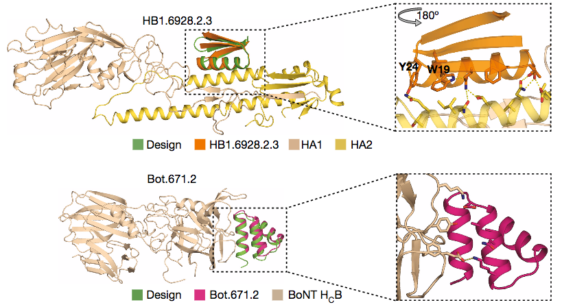
Silva, D. A., Stewart, L., Lam, K. H., Jin, R. & Baker, D. Structures and disulfide cross-linking of de novo designed therapeutic mini-proteins. FEBS J 285, 1783-1785 (2018) doi: 10.1111/febs.14394. PMCID: PMC6001749
Lam, K. H., Qi, R., Liu, S., Kroh, A., Yao, G., Perry, K., Rummel, A. & Jin, R. The hypothetical protein P47 of Clostridium botulinum E1 strain Beluga has a structural topology similar to bactericidal/permeability-increasing protein. Toxicon 147, 19-26 (2018) doi: 10.1016/j.toxicon.2017.10.012. PMCID: PMC5902665
Chevalier, A., Silva, D.A., Rocklin, G.J., Hicks, D.R., Vergara, R., Murapa, P., Bernard, S.M., Zhang, L., Lam, K.H., Yao, G., Bahl, C.D., Miyashita, S.I., Goreshnik, I., Fuller, J.T., Koday, M.T., Jenkins, C.M., Colvin, T., Carter, L., Bohn, A., Bryan, C.M., Fernández-Velasco, D.A., Stewart, L., Dong, M., Huang, X., Jin, R., Wilson, I.A., Fuller, D.H. & Baker, D. Massively parallel de novo protein design for targeted therapeutics. Nature 550(7674):74-79 (2017) doi: 10.1038/nature23912. PMCID: PMC5802399
Yao, G., Lam, K.H., Weisemann, J., Peng, L., Krez, N., Perry, K., Shoemaker, C.B., Dong, M., Rummel, A. & Jin, R. A camelid single-domain antibody neutralizes botulinum neurotoxin A by blocking host receptor binding. Sci Rep. 7;7(1):7438. (2017) doi: 10.1038/s41598-017-07457-5. PMCID: PMC5547058
Yao, G., Lam, K.H., Perry, K., Weisemann, J., Rummel, A. & Jin, R. Crystal Structure of the Receptor-Binding Domain of Botulinum Neurotoxin Type HA, Also Known as Type FA or H. Toxins (Basel) 9, 93 (2017) doi: 10.3390/toxins9030093. PMCID: PMC5371848
Yao, G., Zhang, S., Mahrhold, S., Lam, K. H., Stern, D., Bagramyan, K., Perry, K., Kalkum, M., Rummel, A.*, Dong, M.* & Jin, R.* N-linked glycosylation of SV2 is required for binding and uptake of botulinum neurotoxin A. Nat Struct Mol Biol 23 (7):656-662 (2016) (*corresponding authors) doi: 10.1038/nsmb.3245. PMCID: PMC5033645
Lee, K., Lam, K. H., Kruel, A. M., Mahrhold, S., Perry, K., Cheng, L. W., Rummel, A. & Jin, R. Inhibiting oral intoxication of botulinum neurotoxin A complex by carbohydrate receptor mimics. Toxicon 107, 43-49 (2015) doi: 10.1016/j.toxicon.2015.08.003. PMCID: PMC4658216
Lam, K.H. & Jin, R. Architecture of the botulinum neurotoxin complex: a molecular machine for protection and delivery. Current Opinion in Structural Biology 31:89-95 (2015) doi: 10.1016/j.sbi.2015.03.013. PMCID: PMC4476938
Lam, K.H., Yao, G. & Jin, R. Diverse binding modes, same goal: The receptor recognition mechanism of botulinum neurotoxin. Progress in Biophysics and Molecular Biology 117(2-3):225-31 (2015) doi: 10.1016/j.pbiomolbio.2015.02.004. PMCID: PMC4417461
Lam, T.I., Stanker, L.H., Lee, K., Jin, R. & Cheng, L.W. Translocation of botulinum neurotoxin serotype A and associated proteins across the intestinal epithelia. Cellular Microbiology 17(8):1133-1143 (2015) doi: 10.1111/cmi.12424. PMCID: PMC4610714
Matsui, T.*, Gu, S., Lam, K.H., Carter, L.G., Rummel, A., Mathews, II. & Jin, R.* Structural Basis of the pH-Dependent Assembly of a Botulinum Neurotoxin Complex. J. Mol. Biol. 426(22):3773-3782 (2014) doi: 10.1016/j.jmb.2014.09.009. (*corresponding authors) PMCID: PMC4252799
Lee, K., Zhong, X., Gu, S., Kruel, A.M., Dorner, M.B., Perry, K., Rummel, A., Dong, M. & Jin, R. Molecular basis for disruption of E-cadherin adhesion by botulinum neurotoxin A complex. Science 344(6190):1405-1410 (2014) doi: 10.1126/science.1253823. PMCID: PMC4164303
Lee, K., Lam, K.H., Kruel, A.M., Perry, K., Rummel, A. and Jin, R. High-resolution crystal structure of HA33 of botulinum neurotoxin type B progenitor toxin complex. Biochem. Biophys. Res. Commun. 446(2):568-573 (2014) doi: 10.1016/j.bbrc.2014.03.008. PMCID: PMC4020412
Yao, Y., Lee, K., Gu, S., Lam, K.H. & Jin, R. Botulinum Neurotoxin A Complex Recognizes Host Carbohydrates through Its Hemagglutinin Component, Toxins (Basel) 6(2):624-635 (2014) doi: 10.3390/toxins6020624. PMCID: PMC3942755
Lee, K., Gu, S., Jin, L., Le, T.T.N., Cheng, L.W., Strotmeier, J., Kruel, A.M., Yao, G., Perry, K., Rummel, A.* & Jin, R.* Structure of a Bimodular Botulinum Neurotoxin Complex Provides Insights into Its Oral Toxicity. PLoS Pathog. 9(10): e1003690 (2013) doi:10.1371/journal.ppat.1003690. (*corresponding authors) PMCID: PMC3795040
Zong, Y. and Jin, R. Structural mechanisms of the agrin-LRP4-MuSK signaling pathway in neuromuscular junction differentiation. Cell. Mol. Life Sci. 70(17):3077-88 (2013) doi: 10.1007/s00018-012-1209-9. PMCID: PMC4627850
Gu, S. and Jin, R. Assembly and function of the botulinum neurotoxin progenitor complex. Curr. Top. Microbiol. Immunol. 364:21-44 (2013) doi: 10.1007/978-3-642-33570-9_2. PMCID: PMC3875173
Gu, S., Rumpel, S., Zhou, J., Strotmeier, J., Bigalke, H., Perry, K., Shoemaker, C.B., Rummel, A. & Jin, R. Botulinum neurotoxin is shielded by NTNHA in an interlocked complex. Science 335(6071):977-81 (2012) doi: 10.1126/science.1214270. PMCID: PMC3545708
Zong, Y., Zhang, B., Gu, S., Lee, K., Zhou, J., Yao, G., Figueiredo, D., Perry, K., Mei, L.* & Jin, R.* Structural basis of neuron-specific regulation of postsynaptic differentiation. Gene & Development 26:247-258 (2012) doi: 10.1101/gad.180885.111. (*corresponding authors) PMCID: PMC3278892
Yao, G., Zong, Y., Gu, S., Zhou, J., Xu, H., Mathews, II. & Jin, R. Crystal structure of the glutamate receptor GluA1 amino-terminal domain. Biochem. J. 438(2):255-63 (2011) doi: 10.1042/BJ20110801. PMCID: PMC3296483
Strotmeier, J., Gu, S., Jutzi, S., Mahrhold, S., Zhou, J., Pich, A., Eichner, T., Bigalke, H., Rummel, A.*, Jin, R.* & Binz, T*. The biological activity of botulinum neurotoxin type C is dependent upon novel types of ganglioside binding sites. Mol. Microbiol. 81(1):143-56 (2011) doi: 10.1111/j.1365-2958.2011.07682.x. Epub 2011 Jun 2. (*corresponding authors)
Strotmeier, J., Lee, K., Völker, A.K., Mahrhold, S., Zong, Y., Zeiser, J., Zhou, J., Pich, A., Bigalke, H., Binz, T., Rummel, A.* & Jin, R.* Botulinum neurotoxin serotype D attacks neurons via two carbohydrate-binding sites in a ganglioside-dependent manner. Biochem. J. 431(2):207-16 (2010) (*corresponding authors)
Jin, R.*, Singh, S.K., Gu, S., Furukawa, H., Sobolevsky, A.I., Zhou, J., Jin, Y. & Gouaux E.* Crystal structure and association behavior of the GluR2 amino-terminal domain. EMBO J. 28(12):1812-23 (2009) (*corresponding authors) PMCID: PMC2699365
Kumar, J., Schuck. P., Jin, R. & Mayer, M.L. The N-terminal domain of GluR6-subtype glutamate receptor ion channels. Nat. Struct. Mol. Biol. 16(6):631-8 (2009) PMCID: PMC2729365
Jin, R., Rummel, A., Binz, T. & Brunger, A.T. Botulinum neurotoxin B recognizes its protein receptor with high affinity and specificity. Nature 444:1092-5 (2006)
Jin, R., Clark, S., Weeks, A.M., Dudman, J.T., Gouaux, E. & Partin, K.M. Mechanism of positive allosteric modulators acting on AMPA receptors. J. Neurosci. 25(39):9027-36 (2005)
Jin, R., Junutula, J.R., Matern, H.T., Ervin, K.E., Scheller, R.H. & Brunger, A.T. Exo84 and Sec5 are competitive regulatory Sec6/8 effectors to the RalA GTPase. EMBO J. 24:2064-74 (2005)
Jin, R., Bank, T., Mayer, M. L., Traynelis, S. & Gouaux, E. Structural basis for partial agonist action at ionotropic glutamate receptors. Nat. Neurosci. 6(8):803-10 (2003)

Jahid S, Ortega JA, Vuong LM, Acquistapace IM, Hachey SJ, Flesher JL, La Serra MA, Brindani N, La Sala G, Manigrasso J, Arencibia JM, Bertozzi SM, Summa M, Bertorelli R, Armirotti A, Jin R, Liu Z, Chen CF, Edwards R, Hughes CCW, De Vivo M, Ganesan AK. PMID: 35385746; PMCID: PMC9127750.
Chen P, Zeng J, Liu Z, Thaker H, Wang S, Tian S, Zhang J, Tao L, Gutierrez CB, Xing L, Gerhard R, Huang L, Dong M, Jin R. PMID: 34145250; PMCID: PMC8213806.
Chen P, Lam KH, Liu Z, Mindlin FA, Chen B, Gutierrez CB, Huang L, Zhang Y, Hamza T, Feng H, Matsui T, Bowen ME, Perry K, Jin R. PMID: 31308519; PMCID: PMC6684407.

This new method of drug development relies on the integration of computer modeling and laboratory testing to rapidly generate and evaluate tens of thousands of potential mini-protein binders with varying shapes. This unprecedented scale of de novo protein design was made possible by recent advances in both the Rosetta software suite and DNA manufacturing. Using this method, mini-protein binders can be rapidly programmed to target a range of proteins, including other viruses, toxins, or even tumor-specific markers.
Aaron Chevalier*, Daniel-Adriano Silva*, Gabriel J. Rocklin*, Derrick R. Hicks, Renan Vergara, Patience Murapa, Steffen M. Bernard, Lu Zhang, Kwok-Ho Lam, Guorui Yao, Christopher D. Bahl, Shin-Ichiro Miyashita, Inna Goreshnik, James T. Fuller, Merika T. Koday, Cody M. Jenkins, Tom Colvin, Lauren Carter, Alan Bohn, Cassie M. Bryan, D. Alejandro Fernández-Velasco, Lance Stewart, Min Dong, Xuhui Huang, Rongsheng Jin, Ian A. Wilson, Deborah H. Fuller& David Baker.
In a C. diff infection (CDI), TcdB targets colonic epithelia and binds to what are called Frizzled (FZD) receptors. Researchers in the labs of Rongsheng Jin, PhD, professor of physiology & biophysics from the UCI School of Medicine, and Min Dong, PhD, from Boston Children"s Hospital - Harvard Medical School, found that during this binding process, the toxin locks certain lipid molecules in FZD, which block critical Wnt signaling that regulates renewal of colonic stem cells and differentiation of the colonic epithelium.
"This toxin is indeed very smart. It takes advantage of an important lipid that FZD uses for its own function, to improve its binding affinity and specificity to FZD," said Jin, "However, the need for this lipid also exposes a vulnerability of TcdB that could be exploited to develop antitoxins that block toxin-receptor recognition."
Jin and Dong believe that the novel FZD-antagonizing mechanism exploited by toxin B could be used to turn this deadly toxin into a potential pharmacological tool for research and therapeutic applications, including anti-cancer drugs.

"This is the first time we could directly see the 3D structure of the gigantic TcdB holotoxin at a near atomic resolution," said Rongsheng Jin, PhD, a professor in the Department of Physiology & Biophysics at UCI"s School of Medicine and the senior author in the study. "Interestingly, this toxin shapes like a question mark when viewed from a certain angle, and it has been a major question for us as we seek ways to fight the toxin and CDI."

If not otherwise stated, strains were grown at 20°C as previously described (Brenner, 1974). Strains used in this study were the wild-type Bristol N2, CZ900 syd-2(ju37) (Q397stop point mutation [Zhen and Jin, 1999]; primers 5‘-CACAACGCACCGATCA/3‘-CTTGCATTCGTTCTTTGA), ZM607 syd-2(ok217) (2-kb deletion leading to an early stop after aa 200 [Fig. S2]; primers 5‘-TTGCATCTGCAAAAGAAACG/3‘-GCTCCGAACGAAAGAAGTTG), CZ4601 syd-2(ju487) (R184C point mutation; primers 5‘-AGATGAAGAGATTGTCGAG/3‘-GGTATGTGATGTATTCTCG), VC2392 elks-1(ok2762) (deletion mutation; primers 5‘-CGAATGGAACTCCGAGAACG/3‘-CTTGCACAAGATCCCGATTC), VC223 tom-1(ok285) (deletion mutation; hypomorphic allele) ordered from the Caenorhabditis Genetics Center (College of Biological Sciences, University of Minnesota), and CZ5971 elks-1(js816);syd-2(ju487), CZ5821 elks-1(js816);syd-2(ju37), CZ333 juIs1[Punc-25::SNB-1-GFP] IV, a marker for presynaptic SV clusters in GABAergic motoneurons (Hallam and Jin, 1998), CZ6234 syd-2(ju37);juIs1[Punc-25::SNB-1-GFP], CZ4231 syd-2(ok217);juIs1[Punc-25::SNB-1-GFP], CZ4576 syd-2(ju487);juIs1[Punc-25::-SNB-1-GFP], and GQ707 zxIs6 V [Punc-17::ChR2(H134R)::YFP;lin-15+] expressing a ChR2 (channelrhodopsin2)-YFP fusion protein in cholinergic motoneurons (Liewald et al., 2008). NM2775 elks-1(js816) (a deletion that results in a premature stop at aa 292 [Fig. S2; Deken et al., 2005]; primers: 5‘-CTCACCACAGCTTTAGAATCCG/3‘-CTCACCGCTTCTTTGCTTGCAC) and NM1657 unc-10(md1117) X (8,601-bp deletion that removes the entire UNC-10 coding region; primers 5‘-CGCCCTCTTTCTCTTTCCTTAC/3‘-AGCCTACTAACCCATTTCTCACTG) were provided by M. Nonet (Washington University School of Medicine, St. Louis, MO). J. Kaplan (Massachusetts General Hospital, Boston, MA) provided nuIs152 [Pttx-3::mRFP; Punc-129::GFP–SNB-1]II, which marks presynaptic SV clusters in a subset of dorsally projecting DA and DB motoneurons (Sieburth et al., 2005). GQ751 syd-2(ok217);zxIs6, GQ752 syd-2(ju487);zxIs6, GQ171 syd-2(ju37)X;nuIs152 [Pttx-3::mRFP;Punc-129::GFP-SNB-1]II, GQ172 syd-2(ok217)X;nuIs152 [Pttx-3::mRFP;Punc-129::GFP-SNB-1]II, and GQ173 syd-2(ju487)X;nuIs152 [Pttx-3::mRFP;Punc-129::GFP-SNB-1]II were generated in this study.
For SYD-2 protein quantification, mixed-stage worm extracts were prepared as reported previously (Eimer et al., 2007). In short, six large 9-cm plates containing worms of all stages from egg to young adult were rinsed off and washed two times with M9 buffer, pelleted by centrifugation, and resuspended in 400 µl homogenization buffer including protease inhibitor (cOmplete, EDTA-free Protease Inhibitor Cocktail Tablets; Roche). Samples were frozen at −80°C for 30 min and sonicated, and total protein content was measured. About 50 µg of total protein extracts per strain was loaded and separated on 7.5% SDS-PAGE gel, blotted onto nitrocellulose membrane, and probed against SYD-2 and actin. The polyclonal goat antibody against the N-terminal region of SYD-2 (Santa Cruz Biotechnology, Inc.) and the polyclonal rabbit antibody (Sigma-Aldrich) were used at 1:500 dilutions. Peroxidase-coupled secondary antibodies (donkey anti–goat [Santa Cruz Biotechnology, Inc.]; goat anti–rabbit [The Jackson Laboratory]) were used at 1:10,000 dilutions. The signal was revealed with a camera (LAS-3000; Fujifilm). These experiments were executed at least twice.
The following bacterial expression constructs for GST fusion and His6-tagged proteins were generated using Gateway cloning system (Invitrogen) as described in the manufacturer’s protocols: ELKS-1/pGEX-6P (pCZGY688), ELKS-1/pRSET B (pCZGY659), SYD-2 N (1–517) wild type/pRSET (pCZGY682), and SYD-2 N (1–517) R184C/pRSET (pCZGY683). The SYD-2 fragment (aa 1–515) was cloned between BamHI and SalI sites of a modified pGEX-6P vector to generate SYD-2 N wild type/pGEX-6P (pCZ757) or SYD-2 N R184C/pGEX-6P (pCZ758). SYD-2 91–207 was cloned between EcoRI and BamHI sites of a pMAL-c2 vector to generate SYD-2 91–207/pMAL-c2 or SYD-2 91–207 R184C/pMAL-c2 (gifts from R. Jin, Sanford-Burnham Medical Research Institute, La Jolla, CA). Plasmids were confirmed by sequencing.
Recombinant GST, His6, and maltose binding protein (MBP) fusion proteins were produced in E. coli BL21(DE3) with pGEX-6P, pRSET, and pMAL-c2 plasmids incubating for 16 h at 22°C in the presence of 0.1, 0.5, and 0.3 mM IPTG, respectively. Proteins were extracted by sonication in PBS, pH 7.4, with 0.1% Triton X-100, 1 mM DTT, and protease inhibitors (Roche). For GST pull-down analysis, GST fusion proteins in supernatant were immobilized to glutathione-Sepharose (GE Healthcare), incubated with His6 proteins in supernatants for 3 h at 4°C, and washed four times. Protein complexes were eluted and analyzed by Ponceau S staining and Western blotting using a chemiluminescent substrate (SuperSignal West Femto; Thermo Fisher Scientific) and mouse monoclonal anti-His6 (EMD Millipore) antibody. For gel filtration analysis, MBP-LH1 and MBP-LH1 (R184C) were purified with Amylose resin (New England Biolabs, Inc.), and 40 µg proteins was applied to an HPLC (1100; Agilent Technologies) with PROTEIN KW-803 column (Shodex) equilibrated with 20 mM potassium phosphate, pH 6.8, and 50 mM sodium chloride at a 0.5-ml/min flow rate, detected by a MALS instrument (DAWN HELEOS 8+; Wyatt) and a refractive index detector (Optilab rEX; Wyatt). These experiments were executed twice.
Fig. S1 shows 3D reconstructions of elongated AZ DP from neuron–neuron synapses from tomography and serial thin sections. Fig. S2 shows the gene and protein domain organizations of syd-2 and elks-1 as well as the position of the mutations and deletions used, and protein levels in the respective syd-2 mutants were analyzed by Western blot. Fig. S3 shows the pattern of AZ SV clusters marked by a synaptobrevin-GFP (SNB-1–GFP) fusion protein in cholinergic and GABAergic motoneurons of the different syd-2 mutants. Fig. S4 shows the sensitivity of the different syd-2 mutants to the drugs aldicarb and levamisole. Fig. S5 show a schematic representation of how the length of the presynaptic AZ DP is controlled by the activity of SYD-2 through the recruitment of ELKS-1. Online supplemental material is available at http://www.jcb.org/cgi/content/full/jcb.201302022/DC1.

Quantitative immuno-positron emission tomography imaging of HER2-positive tumor xenografts with an iodine-124 labeled anti-HER2 diabody. Cancer Res. 2005 Feb 15; 65(4):1471-8.
A single treatment of yttrium-90-labeled CHX-A"-C6.5 diabody inhibits the growth of established human tumor xenografts in immunodeficient mice. Cancer Res. 2004 Sep 01; 64(17):6200-6.

Zhang H, H Hu, N Greeley, J Jin, A Matthews, E Ohashi, M Caetano, H Li, X Wu, P Mandel, J McMurray, SJ Moghaddam, S-C Sun, S. Watowich. STAT3 restrains RANK- and TLR4-mediated signalling by suppressing expression of the E2 ubiquitin-conjugating enzyme Ubc13. Nature Communications 2014 Dec 15; 5:5798. doi: 10.1038/ncomms6798. (2014) PMID: 25503582
Chang JH, Xiao Y, Hu H, Jin J, Yu J, Zhou X, Wu X, Johnson HM, Akira S, Pasparakis M, Cheng X, Sun SC. Ubc13 maintains the suppressive function of regulatory T cells and prevents their conversion into effector-like T cells. Nat Immunol. 13: 481-490 (2012) PMID: 22484734
Hu H#, Wu X#, Jin W, Chang M, Cheng X and Sun S-C. Noncanonical NF-κB regulates inducible costimulator (ICOS) ligand expression and T follicular helper cell development. PNAS 2011 Aug 2; 108(31):12827-32. Epub 2011 Jul 18. [* co-first author] PMID: 21768353
Zhang M#,Wu X#, AJ Lee, W Jin, M Chang, A Wright, T Imaizumi, and S-C Sun. Regulation of IKK-related kinases and antiviral responses by tumor suppressor CYLD. J Biol Chem. 283(27): 8621-6 (2008). PMID: 18467330
Reiley WW, Jin W, Lee AJ, Wright A, Wu X, Tewalt EF, Leonard TO, Norbury CC, Fitzpatrick L, Zhang M, Sun SC. Deubiquitinating enzyme CYLD negatively regulates the ubiquitin-dependent kinase Tak1 and prevents abnormal T cell responses. J Exp Med. 204(6): 1475-85 (2007). PMID: 17548520
Jin W, Reiley WR, Lee AJ, Wright A,Wu X, Zhang M, Sun SC. Deubiquitinating enzyme CYLD regulates the peripheral development and naive phenotype maintenance of B cells. J Biol Chem. 282(21): 15884-93 (2007). PMID: 17392286
Babu GR, Jin W, Norman L, Waterfield M, Chang M, Wu X, Zhang M, Sun SC. Phosphorylation of NF-kappaB1/p105 by oncoprotein kinase Tpl2: implications for a novel mechanism of Tpl2 regulation. Biochim Biophys Acta. 1763(2): 174-81 (2006). PMID: 16448710


 8613371530291