
overshot jaw in puppies factory
This website is using a security service to protect itself from online attacks. The action you just performed triggered the security solution. There are several actions that could trigger this block including submitting a certain word or phrase, a SQL command or malformed data.
An overbite might not seem like a serious condition for your dog, but severely misaligned teeth can lead to difficulty eating, gum injuries and bruising, bad breath and different types of dental problems, including tooth decay and gingivitis. Fortunately, there are ways to help fix the problem before it becomes irreversible.
An overbite is a genetic, hereditary condition where a dog"s lower jaw is significantly shorter than its upper jaw. This can also be called an overshot jaw, overjet, parrot mouth, class 2 malocclusion or mandibular brachynathism, but the result is the same – the dog"s teeth aren"t aligning properly. In time, the teeth can become improperly locked together as the dog bites, creating even more severe crookedness as the jaw cannot grow appropriately.
This problem is especially common in breeds with narrow, pointed muzzles, such as collies, shelties, dachshunds, German shepherds, Russian wolfhounds and any crossbred dogs that include these ancestries.
Dental examinations for puppies are the first step toward minimizing the discomfort and effects of an overbite. Puppies can begin to show signs of an overbite as early as 8-12 weeks old, and by the time a puppy is 10 months old, its jaw alignment will be permanently set and any overbite treatment will be much more challenging. This is a relatively narrow window to detect and correct overbites, but it is not impossible.
Small overbites often correct themselves as the puppy matures, and brushing the dog"s teeth regularly to prevent buildup can help keep the overbite from becoming more severe. If the dog is showing signs of an overbite, it is best to avoid any tug-of-war games that can put additional strain and stress on the jaw and could exacerbate the deformation.
If an overbite is more severe, dental intervention may be necessary to correct the misalignment. While this is not necessary for cosmetic reasons – a small overbite may look unsightly, but does not affect the dog and invasive corrective procedures would be more stressful than beneficial – in severe cases, a veterinarian may recommend intervention. There are spacers, braces and other orthodontic accessories that can be applied to a dog"s teeth to help correct an overbite. Because dogs" mouths grow more quickly than humans, these accessories may only be needed for a few weeks or months, though in extreme cases they may be necessary for up to two years.
If the dog is young enough, however, tooth extraction is generally preferred to correct an overbite. Puppies have baby teeth, and if those teeth are misaligned, removing them can loosen the jaw and provide space for it to grow properly and realign itself before the adult teeth come in. Proper extraction will not harm those adult teeth, but the puppy"s mouth will be tender after the procedure and because they will have fewer teeth for several weeks or months until their adult teeth have emerged, some dietary changes and softer foods may be necessary.
An overbite might be disconcerting for both you and your dog, but with proper care and treatment, it can be minimized or completely corrected and your dog"s dental health will be preserved.

This condition is most often spotted at either the first or second puppy checks or between 6 and 8 months of age as the permanent (adult) teeth erupt. Either the deciduous or permanent lower canines occlude into the soft tissues of the roof or the mouth causing severe discomfort and, possibly, oral nasal fistulae.
The fact sheet answers many questions you may have about the cause of this problem and the various treatments available. It is important not to delay treatment of deciduous lower canines as the window of opportunity is only a matter of a few weeks until the permanent canines erupt at 22 to 26 weeks of age. A new problem can then present with bigger teeth causing more damage.
We advise you email us images of the teeth (mouth closed, lips up and side on for both left and right) just a few days before you travel. Things change quickly in growing dogs and it might save you a wasted journey.
This is an inherited condition - an autosomal recessive mutation. Both parents may look normal but carry recessive genes for the condition. When this genetic information is passed onto the litter, approximately one pup in four will be affected, appear abnormal and can pass the genetic information on if bred from. In addtion, two pups in four will carry an abnormal gene from one parent and a normal gene from the other. This pups will look normal but can pass the problem on if bred. Finally one pup in four will not be a carrier of abnormal genes, will be unaffected and cannot pass the trait on to future generations.
If this condition appears in the litter, the most responsible course of action is not to breed from the parents again - either as a pair or individually with others. As there is currently no test to identify this gene, selecting another mate may mean they too are recessive carriers. All the normal looking sibling pups are likely to also carry the recessive genes. It is wise that they too do not contribute to passing the problem back into the breed"s gene pool. In many affected breeds, the gene pool of breeding individuals to select from is very small. If recessive carriers are routinely mating then it is not long before increasing numbers of pups appear with this condition. Over four decades we have monitored the breeds treated here and it is disappointing to note that many previously unaffected breeds are now being seen on a regular basis.
When a pup is treated for this condition we routinely supply the Kennel Club with a Change of Conformation form so they can track the parental origin. We also ask for permssion to send a DNA swab to the Animal Health Trust. This is anonymously evaluated as part of a research programme to identify the exact genetic origin of the condition with the aim of a simple test becoming available to identify recessive carriers. In time this will allow owners of known recessive carriers to select a mate unaffected by the condition.
Owners with young puppies identified with this problem at first presentation are advised to have the deciduous lower canines removed as soon as possible. There are three reasons for this:
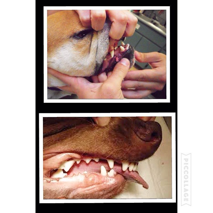
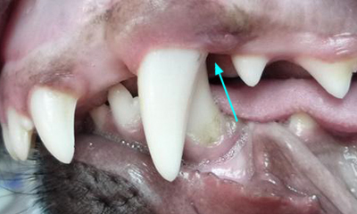
Firstly, and most importantly, these teeth are sharp and hitting the soft tissues of the palate. These pups cannot close their mouth without pain and often hold the mouth slightly open to avoid contact. This is not pleasant. See above for an example of the damage caused to the hard palate by this problem.
Secondly, the growth of the mandible is rostral from the junction of the vertical and horizontal ramus. If the lower canines are embedded in pits in the hard palate, the normal rostral growth of the mandible(s) cannot take place normally due to the dental interlock caused by the lower canines being embedded in hard palate pits. This can cause deviation of the skull laterally or ventral bowing of the mandibles (lower jaws).
Thirdly, the permanent lower canine is located lingual to the deciduous canine. This means that if the deciduous lower canines are in a poor position it is a certainty the permanent teeth will be worse. See the radiograph below. The deciduous canines are on the outside of the jaws and the developing permanent canines are seen in the jaw as small "hats". It is clear that the eruption path of the permanent canines will be directly dorsal and not buccally inclined as is normal.
For these three reasons it is advisable to surgically remove the lower canine teeth as soon as possible to allow maximum time between the surgery and the time the permanent teeth erupt at between 22 and 24 weeks of age. See our file for illustration of removal of deciduous canines.
The deciduous tooth root is three to four times longer than the visible crown and curved - often 2.5cm in length and curved. The root apex is often located below the third lower premolar. See middle and right images below with extracted deciduous tooth laid over extraction site.
The roots are very fragile and will break easily if unduly stressed during removal. A broken root needs to be identified and removed otherwise it continues to form a barrier to the eruption path of the permanent canine and can cause local infection.
The permanent successor tooth is located lingual to the deciduous tooth and wholly within the jaw at this stage. Any use of luxators or elevators on the lingual half of the deciduous tooth will cause permanent damage to the developing enamel of the permanent tooth. See the images below showing canines (and also the third incisor) with extensive damage to the enamel. The radiograph also shows how much damage can occur to the teeth - see the top canine and adjacent incisor. Some severely damaged teeth need to be extracted while other can be repaired with a bonded composite. This damage is avoidable with careful technique using an open surgical approach.
Surgery to remove the deciduous canines may not prevent to need for surgery on the permanent canines but, without it, few cases will resolve if left to nature. Many owners are reluctant to have young pups undergo surgery. Our view is that surgical removal of the lower deciduous canines will not guarantee the problem does not happen again when the permanent teeth erupt but without surgery the chances are very slim.
In a few selected cases - usually only very mild lingual displacement - we can consider placing crown extensions on the lower canines to help guide them into a more natural position. It carries some uncertainly and will not be suited or work in all cases. The images below show crown extensions on a young Springer Spaniel.
Please note that the use of a rubber ball to assist tipping of the deciduous lower canines buccally is not appropriate at this age and will not work - see below.
If the permanent teeth are lingually displaced the pup is usually older than 24 weeks. The trauma caused by the teeth on the soft tissues can be considerable with pain as a consequence.
Do not try ball therapy with deciduous (puppy) teeth. There are two main reasons for this. Puppy teeth are fragile and can easily break. More importantly, the adult canine tooth bud is developing in the jaw medial to the deciduous canine tooth (see radiograph above in the puppy section). If the deciduous crown tips outwards the root will tip inwards. This will push the permanent tooth bud further medial than it already is.
Ball therapy will only work with adult teeth and only in some cases where the lower canines have a clear path to be tipped sideways - laterally - through the space between the upper third incisor and canine. The window of opportunity can be quite short, around 6 weeks, and starts when the lower canine teeth are almost making contact with the hard palate.
If you are considering ball therapy ask your vet their opinion and get them to send us images of each side of the closed mouth from the side with mouth closed and lips up.
The size and type of the ball or Kong is critical. The ball diameter should be the distance between the tips of the two lower canine teeth plus 50%. Therefore if this distance is 30mm the ball diameter is 45mm. If the ball is too small it will sit between the lower canines and produce no tipping force when the pup bites down. Too large a ball can intrude the lower canines back into their sockets.
The owner needs to encourage play with the ball several times a day (6 - 8) or as often as they will tolerate with a short attention span. The ball should be only at the front of the mouth to go any good. If there are no positive results in six weeks a further veterinary evaluation is advised.
These permanent teeth can theoretically be treated by three options. Not all options are available to all cases. These options are described below and are either surgical removal of the lower canines teeth (and possibly incisors also), crown amputation and partial pulpectomy or orthodontics via an inclined bite plane bonded to the upper canines and incisors. The latter option may not be available to all dogs if the diastema (space) between the upper third incisor and canine is too small for the lower canines to move into or if the lower canines are located behind (palatal) to the upper canines.
This is a sterile procedure to reduce the height of the lower canine crown that exposes the pulp. It requires a removal of some pulp (partial coronal pulpectomy) and placement of a direct pulp capping.
This is a very delicate procedure and carries very high success rate (in our hands) since the availability of Mineral Trioxide Aggregate (MTA). We have used it as the material of choice since 2005. The previous agent (calcium hydroxide) was much more caustic and tended to "burn" the pulp. The success rate of MTA treated cases is quoted as 92% in a seminal ten year study based in vet dental clinics in Finland. This compares with 67% when caclium hydroxide was previously the agent. Luotonen N et al, JAVMA, Vol 244, No. 4, February 15, 2014 Vital pulp therapy in dogs: 190 cases (2001–2011).
The intention of the procedure is to keep the pulp alive and allow the shortened lower canines to develop normally and contribute to the strength of the lower jaws.
Radiograph left lower canine before (left) and immediately after (right) surgery. Note the immature morphology of the canine teeth - thin walls and open root apices.
In order to monitor this process of maturation we need to radiograph these teeth twice at 4-6 months post-op and again at 12 -16 weeks post-op. This is a mandatory check. The quoted success reate of 92% implies 8% failure. Half of those to fail in the Luotonen study happened over a year post-op. To ensure any failure of maturity is identified we will not perform this surgery unless the owner agrees to this.
The left radiograph shows the left lower canine immediately after crown amputation and partial pulpectomy. The right radiograph is same tooth 18 weeks post-op. Note the thicker dentine walls, development of an internal dentine bridge between pulp and direct pulp cap and the closed and matured root apex. These three criteria indicate a successful procedure at this stage.
The advantage of this procedure is that the whole of the root and the majority of the crown remain. The strength and integrity of the lower jaw is not weakened by the procedure and long term results are very good due to the use of Mineral Trioxide Aggregate as a direct pulp dressing.
Surgical extraction of the lower canine may seem attractive to clients as the problem is immediately dealt with without the uncertainties of orthodontics and the post-op check that is part of any crown amputation procedure.
However, many owners are concerned (rightly) about the loss of the tooth and the weakness it may cause to the lower jaw(s). It is not our preferred option. This is not an easy surgical extraction and the resulting loss of the root causes a weakness in the lower jaws. This is compounded if both lower canines are removed.
As this is an elective procedure (e.g. sterile) it is possible to use a bone allograft to fill the void created by the loss of the large canine tooth. The graft will promote new bone growth within a few weeks. Grafts can be very expensive as the void to be filled is large. This can increase the cost of the procedure markedly.
In some mild cases of lingual displacement we may be able to use crown extensions for a few weeks. For this treatment we bond composite resin extensions on the lower canines to increase the crown length by around 30%. This allows the lower canines to occupy the correct position and also provides more leverage to tip the crown tips buccally. The crown extensions remain in place for around 2 months and are then removed and the tooth surface smoothed and treated. The major downside is that if the dog damages or breaks them off, you need to return here for repairs. Sticks and other hard objects can easily cause damage and some toys also have to be withdrawn for the treatment period.
Orthodontic tipping as a treatment has the least certain outcome of all three option. It might seem less invasive than surgery but does require very careful case selection and management.
Normally a composite resin bite plane is bonded onto the upper teeth (see below) with an incline cut into the sides. The lower canine makes contact with the incline when the mouth closes and, over time, the force tips the tooth buccally. This takes around four to eight weeks. The lower canine will often migrate back into a lingually displaced position when the bite plane is removed. This can occur if the tooth height of the lower canine is too short (stunted). If the lower canine is not self-retained by the upper jaw when the mouth is shut further surgery may be required.
Orthodontic treatment will also conceal a defect and will not be performed unless the patient is neutered. In addition we have an ethical obligation to inform the Kennel Club of a change in conformation.
The images below show a lingually displaced left lower canine before treatment and after application of a bite plane. The bite plane remains in the mouth as long as it takes for the power of the bite to tip the lower canine into the normal position by pushing it up the incline.
Not all dogs or owners are suited to this. Bite planes can become dislodged if the dog bites a stick or other hard object. Bite planes also need cleaned and adjusted from time to time under sedation or anaesthesia. All of this means more travel and expense for you and more anaesthesia for your pet. It is our view that if a treatment has uncertain outcomes built in it should probably not be used.
Enzo is the Hawthorne Hills Veterinary Hospital Pet of the Month for May. Everyone knows that puppies need vaccines to keep them healthy and protected from diseases. However, it can be easy to underestimate the benefits of thorough and regular examinations when puppies are growing into adulthood. Every breed has special characteristics that make them unique and add to their appeal and sometimes there are physical changes that need to be addressed quickly. For this reason our veterinarians believe in examinations with every vaccine, especially during a puppy’s formative months.
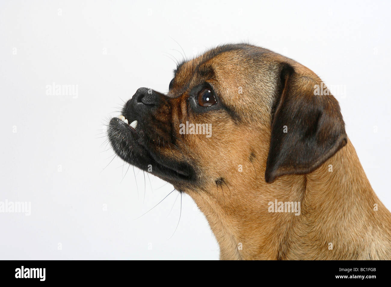
Enzo is a short-haired Havanese and he was born with his lower jaw shorter than the upper jaw. This is called an Overbite, also referred to as an Overshot Jaw, a Parrot Mouth or Mandibular Brachygnathism. This malocclusion is a genetic change and can be seen in a number of breeds, oftentimes collie related breeds and dachshunds. Occasionally this change happens because of differences in the growth of the upper and lower jaws, and in many cases it doesn’t cause any significant problems other than cosmetically.
Dr. Robin Riedinger evaluated Enzo at his first visit when he was just 11 weeks of age and while the lower jaw was too short, there was no evidence of damage and no indication that this was causing a problem for Enzo. When there is abnormal occlusion of the teeth, it is important to monitor closely for trouble caused by the teeth being aligned improperly. Malocclusions can lead to gum injuries, puncturing of the hard palate, abnormal positioning of adjacent teeth, abnormal wear and bruising of the teeth, permanent damage and subsequent death of one or more teeth, and in the long run, premature loss of teeth. Some malocclusions can be severe enough to interfere with normal eating and drinking.
Within three weeks, when Enzo was only 3.5 months old, it was clear that our doctors would need to intervene. The left and right sides of Enzo’s upper jaw (maxilla) were growing at different rates because the lower canine teeth were being trapped by the upper canine teeth. This is called Dental Interlock. Because the teeth are ‘locked’ in place, the lower jaw cannot grow symmetrically and this creates a number of other problems. Early intervention is critical.
The solution for Dental Interlock is to extract the teeth from the shorter jaw; in this case, the lower ‘baby’ canines and thereby allow the lower jaw (mandible) to grow in the best way possible. This procedure is most effective when the Dental Interlock is discovered early and the extractions are performed quickly. In some cases, this can be as early as ten weeks of age. Dr. Riedinger consulted with a local veterinary dental specialist to confirm the treatment plan and to get advice on extracting the deciduous teeth without damaging the developing adult canines. Dental radiographs are essential to proper extraction technique and also to ensure that there are no other abnormalities below the gumline.
You can see how long the roots of the deciduous ‘baby’ teeth are. During normal growth, the body will begin to resorb the roots, making them loose, and allow them to fall out as the adult tooth begins to emerge. When we need to remove the deciduous teeth before they are loose, it can be quite tricky to remove the tooth carefully without breaking it and without injuring the adjacent teeth.
Once extracted, each deciduous canine tooth was about 2 centimeters long; the roots were about 1.5 centimeters. Many people are surprised to learn that the root of a dog’s tooth is so large – 2/3 to 3/4 of the tooth is below the gumline. This is one reason why it is so important to use radiographs to evaluate teeth on a regular basis, not just in a growing puppy. Adult teeth can, and frequently do, have problems that are only visible with a radiograph.
Enzo came through his procedure extremely well. He was given pain medications for comfort and had to eat canned foods and avoid chewing on his toys for the next two weeks to ensure that the gum tissue healed properly. As he continues to grow we will be monitoring how his jaw develops and Dr. Riedinger will also be watching the alignment of his adult canine teeth when they start to emerge around six months of age. Hopefully this early intervention will minimize problems for Enzo in the future.

Generally, there are not a lot of problems that occur to the baby teeth. They aren’t without issues, however. Thankfully their short existence means that there’s not a lot of chance for something to go wrong with them.
The most common problem we see with puppy teeth is that sometimes they do not fall out. This is called retained deciduous teeth. Puppy teeth being deciduous should fall out and be replaced by the permanent teeth. Sometimes the roots of the baby teeth do not disappear, and instead of the deciduous tooth being replaced by the permanent tooth, the two teeth are crowded together trying to fit in the mouth in the same position. This results in two problems:
This is more common in the canine teeth and occurs more in smaller breeds of dogs. If you notice that this is occurring with your dog, have them visit their vet. We diagnose retained deciduous teeth if we see the new adult tooth just erupting through the gum (not fully erupted) and the deciduous tooth is firm to touch (not about to fall out). These retained teeth should be extracted completely (with the root) as soon as they are noticed. Leaving them for too long to “give them time to fall out” unfortunately in most cases causes the adult tooth to become fixed in the incorrect position. It is not an uncommon scenario for these retained teeth to be removed at desexing which often is done around 6 months of age, just at the time when the deciduous teeth should have fallen out.
A not uncommon problem seen in puppies is broken teeth. As with adult dogs, puppies that do break their teeth do not always clearly tell us that there is a problem. The broken tooth is a problem, even if the pup is not indicating any outward signs. A broken tooth, in adults also, always goes on to develop an abscess at the tooth root. The infection that develops is a problem because at the root tip, where the infection goes, is a developing adult tooth. This developing tooth can be damaged by the infection and inflammation that occurs. So, in conjunction with the pain that it also causes, these fractured teeth should be removed when they are noticed.
With much more crossbreeding happening these days, we have seen a rise in jaw problems in dogs. This will often become apparent in the puppy. The top (maxilla) and the bottom (mandible) jaws are under control from two different sets of genes, so when we see breeding between different head shaped dogs we sometimes see the two jaws at different positions.
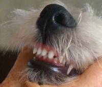
The most common problem we see in jaw development is an undershot jaw. This is also called prognathism or a Class 3 Malocclusion. This is where the mandibles are relatively too long for the maxilla. A classic breed to suffer from this problem is the Boxer or a Shih Tzu. This malocclusion causes the teeth to not line up properly. As they are designed to damage tissue, any tissue, they do! In dogs with an undershot jaw it is not uncommon for the upper incisors (the front teeth) to touch or dig into the bottom jaw, behind the lower incisors. Although this is arguably cute, the teeth over time can cause a great deal of discomfort and even end up damaging the bone and teeth of the lower jaw.
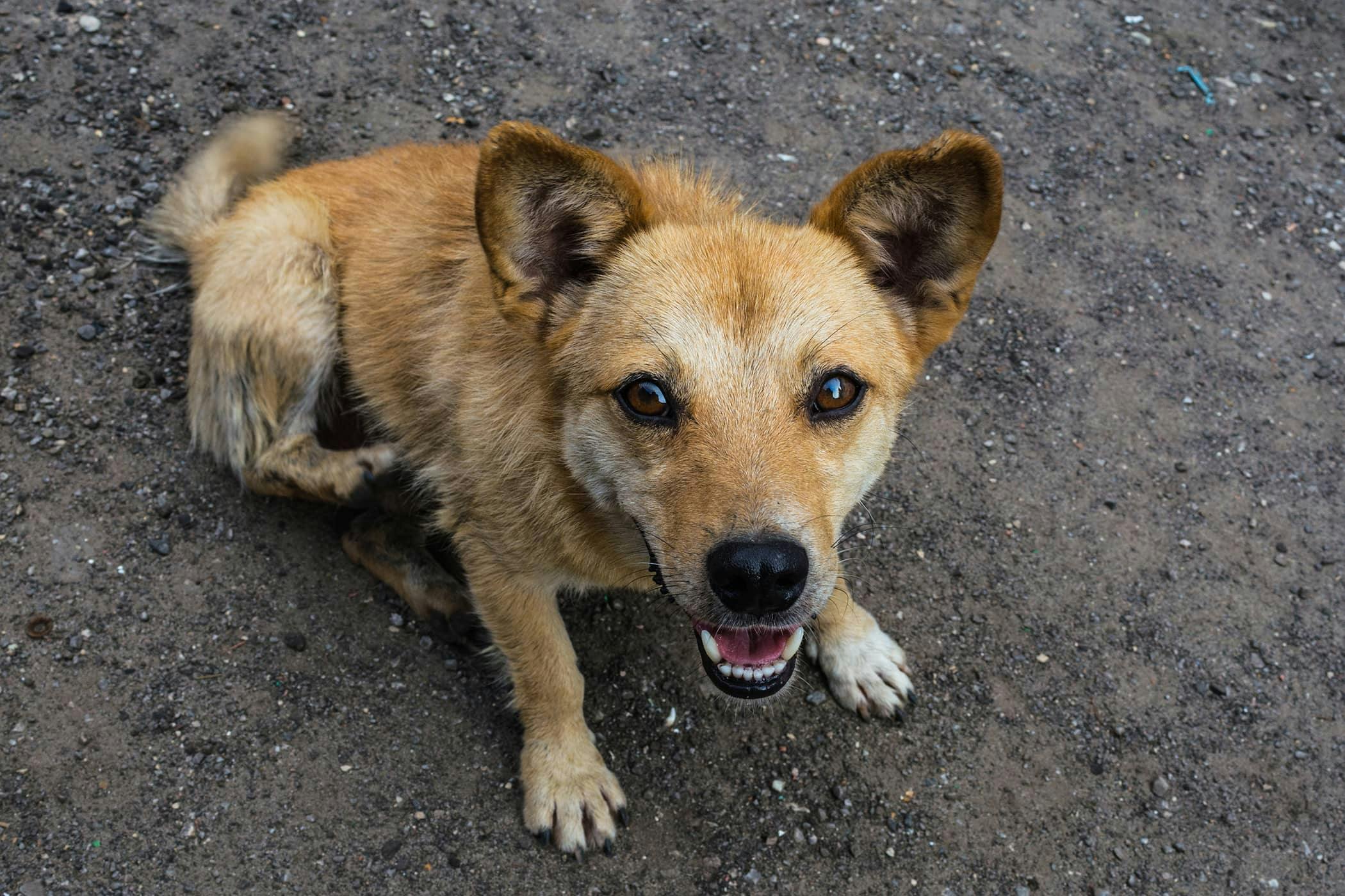
The opposite problem which we are seeing more and more of is an overbite. Also, called brachygnathism or a Class 2 Malocclusion. This is where the top jaw is relatively too long for the mandibles. This can result in a much bigger problem for those dogs affected. Even as puppies the lower deciduous canines can be in such a position that they strike the roof of the mouth. Anyone who has played with puppies knows that these are sharp and painful teeth. Every time these puppies close their mouths it hurts. This also will occur with the following adult canine teeth too. However, they are much larger and can cause much more of a problem with the dog’s mouth.
In puppies with an overbite, it may be necessary to remove the lower deciduous canines early (12 weeks of age) to stop them being in pain until those teeth fall out. If the adult teeth are in a similar position (which they normally are) then they can cause fistulae (holes) to form in the roof of the mouth. These teeth should be examined by a Vet and plans put in place to resolve the problem.
The very best thing we can do to maintain good dental health in our dogs is to brush their teeth. This needn’t be a difficult job, or an upsetting fight that happens every evening (yes you should brush once a day!). If we institute a routine and get the puppy used to having its teeth brushed daily from a young age, everyone involved gets used to the idea quickly. Start with getting the pup used to having your fingers poke around in its mouth, up and under the lips and cheeks. After a few days of this, move onto a finger toothbrush. These should be available at your vet, along with some Pet safe toothpaste. Apart from tablets, everything we put in a pup’s mouth is swallowed, so adult human toothpaste, which must be spat out, is not healthy for dogs. Once the pup is used to daily brushing with a finger toothbrush, add some of the pet friendly toothpaste and continue! The biggest tip for those wanting to keep their pets mouth as healthy as possible – Don’t open their mouth. I even recommend gently holding the mouth closed whilst your finger (with a toothbrush on it) gently moves in a back and forth motion, cleaning the teeth under the lips and cheek.
Hell its my $0.02c and your dog, do what you want. Go look at a heap more puppies and wear out that "Oh so cute must have to hold and cuddle" emotion that puppies invoke and think with your long term brain rather than your paternal desire.
ninja edit: +1 pound puppy, have had a few. Now have Pug. Ultimate kids dog. Did lots of research and bought from very reputable breeder and not factory or backyard where problems end up enhanced.
damn double ninja edit with a twist. My first GF"s mum, y-e-a-r-s ago bred Cav"s. Bought them in from OS to expand bloodlines etc. Learnt a lot about the problem ones as she was bought lots for advice. Daughter was awesome GF except wanted ring and babies @ 18! Aaaaaagh! And I ran. Once they start with an issue they compound with age. This includes experience with Bichons (mops) and Cairn Terriers
Their premolars erupt around 5 to 6 weeks of age. Puppies do not have molars — that really big tooth near the rear of the mouth you probably think is a molar is called the carnassial tooth, and it is actually a premolar.
How to Alleviate Your Puppy’s Gum PainAs the teeth are coming in, your puppy’s gums may hurt. You can help by giving him chew toys in a variety of textures. A toy that can be soaked in or filled with water and frozen will provide
Make sure whatever you give him does not resemble anything of yours that you don’t want him to chew — this means no old shoes! Don’t give him anything that resembles something he can find around the house, either: no socks and no stuffed animals (if you have children who collect them). What your puppy learns to chew on at an early age will tend to be what he looks to chew on for the rest of his life.
Even at this early age, you may notice occlusion problems, or issues with how the upper and lower teeth fit together. Ideally, for most breeds, the upper incisors fit snugly just in front of the lower ones, and the lower canine is just in front of the upper one. In some flat-faced breeds, it’s normal for the jaw to be undershot, with the lower incisors in front of the upper ones, and in some puppies, there may be a small gap between the upper and lower incisors. This very often improves on its own by adulthood.
But in other puppies, the upper jaw may jut out well beyond the lower jaw, and the upper canine tooth may be placed in front of the lower canine tooth. This is an abnormal bite that probably will not get better. As these puppies mature, you must make sure that the short lower jaw, which narrows toward the end, is not so short and narrow that the lower canines jab into the upper gums or roof of the mouth (a condition called base-narrow canines). Resolution of this issue depends
By 3 to 4 months of age, the baby incisors and canines are replaced by permanent ones, followed by the permanent premolars at 4 to 5 months of age. The molars come in around 4 to 6 months of age. The adult dog normally has 42 teeth.
When to Consult Your VeterinarianRetention of baby teeth is a common problem; this happens if the permanent tooth bud doesn’t grow immediately beneath the baby tooth, so the roots of the baby tooth aren’t reabsorbed as they normally are. This happens most often with the canine teeth. If the baby tooth stays there for more than a week it can interfere with the puppy’s occlusion, especially if he’s a toy dog, so you should consult your veterinarian if you suspect this is happening.
Sometimes a baby tooth just remains in place, with no visible permanent tooth. Never have a retained baby tooth pulled without first checking to make sure a permanent tooth is ready to take its place. Sometimes, in toy breeds especially, the permanent tooth never develops and the baby tooth is the best you’ll get!
As your puppy grows he’ll need more chewing toys — even once he’s through teething. Assemble a group of dog toys and only let your puppy have a few at a time, rotating them every few days so he has the excitement of new toys. Be sure to include some interactive toys, such as those he must work at in order to extract food. You can fill these with kibble, soft cheese, canned dog food or peanut butter and then freeze them to make them last even longer. Some toys dispense kibble a piece at a time as the toy is rolled. With luck, your dog will prefer these fancy toys to your fancy belongings.
Just like human babies, puppies have baby teeth that fall out. Most puppies are born without teeth and go through a process known as puppy teething. From birth to six months, sharp puppy teeth erupt from the gums in the jaw in a predictable timeline. Puppies go through teething stages during the development of their teeth, including sore gums, and eventually—the eruption of 28 baby teeth. During teething, puppies may target all kinds of unexpected objects to gnaw and chew on, like baseboards and shoes, to relieve the discomfort. However, most dogs never outgrow the urge to chew. Pet owners are encouraged to learn about how their puppy"s teeth grow in so they can best handle their dog as it ages.
In the front of the mouth, narrow-edged teeth known as incisors will begin to emerge. The incisors are the first to appear at about two to three weeks of age. Puppies have six incisors on both the top and bottom jaw.
Premolars and molars also begin to grow behind canines (the pointed teeth between the incisors and premolars) at three to six weeks of age, with three on the top and bottom of each side. Four needle-like canines appear at age four weeks and frame the incisors, one on each side, top, and bottom.
The last molars appear by six to eight weeks of age. At about eight weeks, the puppy’s permanent teeth begin pushing out deciduous or "milk teeth." The roots of the baby teeth are absorbed by the body, and in most cases, milk teeth simply fall out.
When the deciduous teeth don"t fall out on time, puppies may appear to have a double set of teeth. Retained baby teeth should be extracted by a veterinarian so that permanent teeth have room to grow. Sometimes, a crowded mouth pushes teeth out of alignment, resulting in difficulty eating or poor dental hygiene (which can lead to periodontal disease).
Breeders often let their puppies go to their new owners" homes around eight weeks. Baby teeth will begin to shed, and permanent adult teeth will start to come in. This process is painful for dogs, so providing puppy safe chew toys is recommended. This is a good time to socialize your dog more, look and touch the inside and outside of its mouth, and prepare for teeth brushing.
At this point, all puppy teeth should be gone, and adult teeth emerge. If there are any baby teeth left, let your vet know so it can be removed. Permanent teeth replace the milk teeth tooth-for-tooth and add four premolars and 10 molars. Most pups will have 42 permanent teeth in place by about seven months of age.
While it can vary somewhat between breeds, there is a progression you can expect as your puppy develops new teeth. It"s important to begin handling your puppy"s mouth while it"s young so you can periodically check for any potential tooth problems. Any type of facial swelling, changes in eating habits, unexpected night awakenings, or rubbing of the face are signs of possible oral discomfort. You"ll want to take your pup to the veterinarian if you see:
Spots of blood on your dog"s toys, brown tartar on the teeth, or gums that are bleeding, inflamed, and/or sore. These are common symptoms of periodontal disease, a large oral issue for dogs.
Crooked teeth or malocclusion (misalignment of the upper and lower jaw). While some breeds have a trademark bite, unusual ones could cause chewing issues.
Schedule a visit with your veterinarian for an initial dental exam for your puppy. This examination will include a look at the teeth, gums, and oral cavity. Ask your vet to demonstrate how to clean your pup"s teeth. This way, you"ll know what brushes, toothpaste, and techniques to use.
Get your pup used to the idea of tooth brushing around six months when its adult teeth start to come in. Regular brushing will prevent plaque, stinky breath, disease, and other medical problems. It"s ideal to brush your puppy"s teeth daily, but once or twice a week will work.
Different kinds of teeth serve various functions, based on the position of the mouth and the shape of the tooth. With some breeds, the shape of the jaw impacts how each type of tooth functions. Most dogs have V-shaped upper and lower jaws which allow the mouth to be opened very wide for grasping and capturing prey—or grabbing and holding toys during play. There are several ways dogs use their teeth:
Dogs have eight premolars in the upper jaw and another eight in the lower jaw. They also have four molars in the top and six in the bottom. The extra molars are designed to crush and are used to process vegetable foods and bones.
Dogs have specialized carnassial teeth composed of premolars and molars. As they pass each other during the mouth"s closure, these teeth act like scissors. The carnassial teeth are innovations of the carnivorous animal that requires shearing action to process flesh.
When the mouth is closed, dogs should have a normal "bite." This is very important so that dogs can eat and use their mouth normally (and is judged accordingly in show dogs). A normal bite looks like this:
Malocclusion refers to the abnormal "bite" or fitting of these teeth. Malocclusion can be normal for certain dog breeds due to differences in the shape of the jaw and mouth. For instance, the flat-faced (brachycephalic) dog breeds like Bulldogs have a normal malocclusion because their lower jaw is longer than the upper. However, this allows the teeth to fit incorrectly, which can cause mouth damage as the dog chews. Thus, a veterinarian or veterinary dentist with orthodontic correction should be aware of malocclusion.

Undershot is a class III malocclusion that is also referred to as mandibular prognathism, maxillary brachygnathism, mandibular mesioclusion, or an underbite. This malocclusion is characterized by a shorter upper jaw and a longer lower jaw, resulting in lower teeth that are in front of the upper teeth. While this condition is normal for some breeds, such as Bulldogs, in many breeds it is unusual. An undershot jaw occurs when the lower jaw grows faster than normal and becomes longer than the upper jaw, and is usually evident around 8 weeks of age in puppies. This misalignment can cause soft tissue trauma, such as to the lips. When the incisors meet instead of fitting next to each other, it is called a level bite. When the malocclusion causes the lower incisors to be placed in front of the upper incisors, it is called a reverse scissors bite.
The cause of overshot and undershot jaws in dogs relate to the increased or decreased rate of growth of the upper and lower jaws in relation to one another. This can occur due to a: Genetic disorder Trauma; Systemic infection ;Nutritional disorder; Endocrine disorder; Abnormal setting of puppy teeth; Early or late loss of puppy teeth.
After a quick physical exam, your vet may have to sedate your dog in order to perform a thorough oral exam. This will assess your dog’s skull type and teeth location in relation to the teeth on the opposite jaw. Often, the placement of the upper and lower incisors in relation to one another can determine what type of malocclusion your dog has. Your vet will note any areas of trauma due to teeth striking those areas, and any cysts, tumors, abscesses, or remaining puppy teeth that may be present. A dental X-ray can also help to assess the health of the jaws and teeth. These diagnostic methods will lead to a diagnosis of an overshot or undershot jaw in your dog.
Treatment of a jaw misalignment will depend on the severity of the condition. If your dog has a misalignment, but can still bite and chew food without problems, no treatment may be needed. If the misalignment is caught early in a puppy’s life, it may only be temporary and may correct itself over time. However, there are times when intervention may be needed. If your puppy’s teeth are stopping the normal growth of his jaws, then surgery to remove those puppy teeth may be performed. This may allow the jaws to continue to grow, but will not make them grow. For older dogs who are experiencing pain and trauma due to misaligned jaws and teeth, oral surgery is generally performed to extract teeth that are causing trauma, to move teeth so that they fit, or to create space for a misaligned tooth to occupy. Other therapies include crown reductions or braces.
If your dog is genetically programmed to have an overshot or undershot jaw, intervention can help, but will not slow or stop the abnormal growth of either jaw. Prevent jaw misalignments in puppies by not breeding dogs who have overshot or undershot jaws.

The goals and purposes of this breed standard include: to furnish guidelines for breeders who wish to maintain the quality of their breed and to improve it; to advance this breed to a state of similarity throughout the world; and to act as a guide for judges.
Any departure from the following should be considered a fault, and the seriousness with which the fault should be regarded should be in exact proportion to its degree and its effect upon the health and welfare of the dog and on the dog’s ability to perform its traditional work.
The Caucasian Ovcharka, also sometimes known as the Caucasian Mountain Dog, is a guardian breed from the Caucasus Mountain area. The breed"s origin is shrouded in antiquity. Some claim the breed is a domestication of the wolves of this region, others that the breed developed from Mastiff-Spitz crosses. Some experts contend that the breed naturally developed from a group of sheepdogs that migrated to the Caucasus from Tibet. More recent archaeological findings point to breed origins in Mesopotamia.
What is known for certain is that herdsmen in the mountains of Georgia, Armenia, Azerbaijan, Daghestan, and surrounding countries, and the steppe regions of the northern Caucasus, have for centuries depended on the Caucasian Ovcharka to guard flocks and villages. Legends are written of the breed"s faithfulness, protectiveness, and ferocity when called upon to defend. Type varies geographically throughout the mountain range, and also varies according to the purpose for which the dogs were used. Generally, dogs of the trans-Caucasus regions are more massive, while those found in the steppe regions have a somewhat rangier build, are leggier, and are often short-coated. Modern breeding conforms to a single standard.
The former Soviet government developed state kennels and used the breed for guarding factories and government facilities throughout the former U.S.S.R. In the United States, Caucasian Ovcharka have earned a reputation as trustworthy service dogs.
The Caucasian Ovcharka is a powerful, athletic dog, strongly muscled, and heavily boned in proportion to height. The head is large, wedge-shaped, and tapers slightly to a blunt muzzle with high-set hanging ears, which may be cropped, and deep-set, oval-shaped eyes. The thick tail hangs down to the hock but may be carried above the back as a sickle-shaped hook or ring when the dog is excited or moving. Three coat lengths are accepted, all double-coated and thick. Coat colors include shades of agouti gray, fawn, and reddish, with white markings and often a dark facial mask. Solid white dogs with dark pigmentation occur occasionally in the breed. Gender differences are well expressed in this breed. Males are more massive and more powerful; females are smaller and lighter in build. Honorable scars resulting from field work are not to be penalized.
Caucasians are spirited, intelligent, strong-willed guardian dogs. While gentle and demonstrative with family members, the Caucasian’s active defense reaction and strong territorial instincts make this breed very suspicious of strange people or dogs. They are steady and even tempered but will protect their flock, family, and property from danger - real or perceived - with lightning-quick speed. Caucasian Ovcharka have keen senses, so they are very alert and good trackers. Any change in their surroundings can result in warning barks and growls, particularly at night. The breed is slow to mature and headstrong. Socialization and patient training techniques can be used to temper the Caucasian Ovcharka’s characteristic suspicion and aggressiveness toward strange people and dogs, resulting in a mature dog with good judgment. This breed is hardy and able to adapt to a wide range of climates.
The head is large, with a broad skull and strongly developed cheek bones. Viewed from the top, the gradually tapering skull and muzzle form a one-piece, blunt wedge shape. The stop is slightly defined and not abrupt. The width of the head is emphasized by dense coat that stands away from the sides of the jaws. The male head is more massive than the female head, which is more refined.
The muzzle is shorter than half the length of the head, but powerful and well filled in under the eyes. It tapers slightly to the nose. The topline of the muzzle is parallel to the topline of the skull. The blunt end of the muzzle is formed by thick, dry lips tightly covering a powerful lower jaw. Lip pigment is black.
The jaw provides ample space for a full complement of large, evenly spaced, white teeth meeting in a scissors bite. The line of the incisors is straight and perpendicular to the outside lines of the jaw. Canines are large and long.
Cropped or uncropped. Uncropped, the ears are high set, triangular-shaped, and hang tight to the head. The outer margin of the ear should not be located below the level of the eyes. The ears may be cropped in what is known as a “shepherd’s crop.” The ear flap is removed horizontally and bluntly, close to the head.
Viewed from the front, the forelegs are straight, well-boned, and set parallel and well apart. The length of the front leg (measured from point of elbow to the ground) should be slightly more than one-half of the dog’s height (measured at the withers). The pasterns are short, strong, and, when viewed from the side, slightly sloping. Circumference of the pasterns ranges from 5½ to 6¾ inches in mature males and from 5 to 6 inches in mature females.
The proportion of the length of body vs. the height at the withers is 100:108. The ribs are well sprung and let down to, or slightly below, the elbows. The chest is broad and deep. The line of the back inclines very slightly downward from broad, muscular, prominent withers to a strong, broad back with a straight upper line. The loin is short, broad, and slightly arched. The croup is broad, long, muscular, and nearly flat. Tuck-up is moderate. The skin is thick and elastic.
The rear legs are well-boned and moderately angulated at stifle and hock joints. The hocks are strong, broad, and well let down. Viewed from the rear, the rear pasterns are parallel to each other. From the side, they are perpendicular to the ground. When standing normally, the rear legs are spaced moderately apart and positioned so that a line dropped from the point of buttocks to the ground would fall through the center of the point of hock and the rear pastern.
The tail is set on high. When the dog is in repose, the tail just reaches to the hock, with the bottom third of the tail forming a hook. When the dog is in action or excited, the tail is carried as a sickle-shaped hook or ring above the level of the back. Docked tails are permitted.
The Caucasian Ovcharka has a double coat consisting of longer, coarse outer guard hairs and dense undercoat made up of soft, fine hair. Coat on the muzzle, forehead, and the front of the legs is short and smooth. Longer coat on the cheeks and the backskull stands away from the body and contributes to the bear-like appearance of this breed. Three types of coat lengths are accepted without preference:
Long coat. The hair of the outer coat is very long, forming a “mane.” Extensive feathering on the hind legs gives the appearance of long, silky “pants.” The long hairs feathering the tail on all sides makes it look thick and fluffy.
The following are acceptable colors and markings: Agouti gray - dark, light, silver, reddish, or yellowish - with or without white markings; White, cream, fawn or reddish fawn, tan or reddish tan, fulvous, with or without white markings; Brindle; Piebald; or White with gray patches.
Disqualifications: Solid black (defined as black to the skin with no shading, such as in the Newfoundland). Black and Tan (defined as black and tan like the Rottweiler). Solid chocolate (defined as any shade of solid brown without lighter undercoat or sable overlay, like a Grizzly bear).
Minimum height at maturity, measured at the withers, is 25½ inches for males and 24½ inches for females. Dogs over 27¼ inches and bitches over 25½ inches are preferred. Weight should be in proportion to the height, giving a balanced, imposing appearance.
At the trot, the Caucasian moves freely with strides of moderate length, usually unhurried. The back remains level, and the front and rear legs on each side move in a parallel fashion. The front and rear pasterns flex freely. The back and loin are elastic and springy. As speed increases, however, the width between the legs decreases and the tendency to single track increases until the dog breaks into a heavy, lumbering gallop.
This website is using a security service to protect itself from online attacks. The action you just performed triggered the security solution. There are several actions that could trigger this block including submitting a certain word or phrase, a SQL command or malformed data.
It is very important for you to inspect a puppy for signs of illness, no matter what the seller tells you. Even with a written health guarantee.Once you fall in love with your new puppy, if he or she is sick or even passes away, that health guarantee is not going to make you feel much better.
Important – if 1 puppy in a litter seems to be ill, we advise to not purchase any puppy from that litter. Other pups may just not be showing symptoms yet. Therefore, use this guideline to look at all of the puppies in the litter…before you go on to the next step that brings you closer to choosing a puppy.
On an 8-week Maltipoo puppy, the hair will still be considered a “puppy coat.” Compared to the adult Maltipoos that you see, it will be shorter and finer, so do not worry if the coat is not as full as the puppies’ parents. This said, of course bald patches or hair loss is a huge issue with a pup.
Clear discharge may be allergies. Many dog do have allergies, this is up to you to decide if you want to obtain a pup with signs of allergies, which can be very frustrating (and sometimes expensive) to deal with.This said, allergies can
be specific to the pup"s environment or to the food that he is eating.Do keep in mind that even if you choose a healthy puppy, once he or she is home with you, allergies may develop to your laundry detergent, the dog shampoo
Open each puppy’s mouth and check his teeth. The gums should be a healthy pink and the teeth should be white. There should be no bad breath issues. The most that you should smell is the lingering scent of puppy food.
This is because both the Maltese and the Poodle have a scissors bite. An undershot or overshot bite, when noticeable in a young pup, can turn into a severe problem as he matures.
If you see worms moving around this area, inform the breeder. This type of worm requires three days of continuous worming to remove them. They are associated with fleas and will hatch out in another two weeks. Do not obtain a puppy with worms, as this is a bad sign – the breeder should have had the puppies de-wormed.
Stools should be firm and not runny. Take a close look to see if you spot any worms. Runny stools or any worms is a big warning sign – do not get the puppy.

An underbite is when the lower jaw juts out from underneath the upper jaw. In some dog breeds, this is fairly common and is often an identifying trait of a breed. However, it can create some problems for the dog over time. Here’s a list of dogs that are frequently born with an underbite, as well as some ideas about whether or not you should correct this.
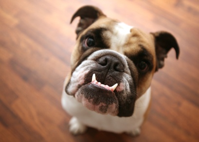
Bulldogs are famous for underbites, and it comes with the territory of the breed. It doesn’t matter if it’s an English bulldog, a French bulldog, or an American bulldog. They all have an underbite as a characteristic of their head and jaw structure. Their recessed nasal passages often push the upper jaw back, causing the lower jaw to jut forward a bit. As long as it doesn’t cause major chewing problems or create major dental issues in your bulldog, you don’t really have to correct it.
Many of the small Asian lap dogs suffer from an underbite, and the Pekingese is no exception. The narrow, upturned nose of the Pekingese shares a trait with the bulldog family in that the upper jaws follow the flattened noses. The Pekingese has a v-shaped lower jaw that doesn’t always meet the unusual shape of the upper jaw and nose area. Subsequently, you will see these little dogs with one or more teeth sticking out of the bottom.
Here is another little Asian lap dog that frequently has an underbite. However, the Shih Tzu should have the underbite corrected if possible. It interferes quite a bit with the little dog’s ability to bite and chew. Their little razor-like teeth can cause dental and oral problems too. The only upshot is that it is easier to brush the Shih Tzu’s exposed lower teeth and prevent tooth decay.
Pugs are often seen with an underbite too. For them, it’s a cute breed trait that doesn’t typically cause them much harm. They have more trouble breathing than they do biting and chewing. Most dogs that have these flattened facial features and smushed-in noses are the ones with an underbite. Pugs are bred to have these facial features, and thus they have underbites.

This website is using a security service to protect itself from online attacks. The action you just performed triggered the security solution. There are several actions that could trigger this block including submitting a certain word or phrase, a SQL command or malformed data.

The primary function of the mouth is to obtain and introduce food into the digestive tract. Some of its additional functions include communication and social interaction, grooming, protection, and heat regulation (particularly in dogs). Picking up food, chewing, and swallowing require a complex interaction of the muscles of the jaw, the teeth, the tongue, and the upper throat. When any of these functions becomes compromised through disease or trauma, malnutrition and dehydration may result. A complete oral examination should be a part of your animal’s physical examination, because oral diseases are most effectively treated with early diagnosis. Otherwise, many will remain hidden in the mouth and progress to an advanced stage. Oral Inflammatory and Ulcerative Diseases
Gum diseaseGum DiseaseDieffenbachia may cause oral inflammation and sores if chewed. Chronic kidney failure can cause inflammation and sores in the mouth.
Signs vary with the cause and extent of inflammation. Loss of appetite may be seen. Bad breath and drooling are common with mouth inflammation, tongue inflammation, and sore throat. The saliva may be tinged with blood. The animal may paw at its mouth and, due to pain, resent or resist any attempt to examine its mouth. Lymph nodes in the region may be enlarged. Canine Stomatitis
Canine stomatitis involves inflammation of the mucous membranes of the mouth. Signs include severe gum inflammation, receding gums in several sites, and large sores on the mouth surface near the surfaces of large teeth. The problem commonly affects Greyhounds, but it has also been seen in Maltese, Miniature Schnauzers, Labrador Retrievers, and other breeds. The characteristic feature is the contact ulcer or sore that develops where the lip contacts the tooth surface, most commonly on the inner surface of the upper lip next to the upper canine and carnassial teeth (also called the 4th premolar). These abnormalities have also been termed “kissing ulcers” because they are found where the lips “kiss” the teeth. Blood tests and tissue samples can rule out other causes of stomatitis (such as advanced kidney disease).
The cause of this disease is an immune system dysfunction that results in an excessive inflammatory response to dental plaque. For this reason, thorough plaque control through professional cleaning and excellent home oral hygiene (including twice daily tooth brushing) may resolve the problem. Supplemental antibacterial measures, such as topical chlorhexidine rinses or gels, may be prescribed by your veterinarian. In severe cases, topical anti-inflammatory preparations may provide comfort. Discomfort caused by the ulcers can make it difficult to brush your pet’s teeth and give oral medications. If discomfort is severe and you are unable to brush the teeth, extraction of the adjacent teeth may be necessary to remove the contact surfaces on which plaque accumulates. Although extraction may aid in control of the sores, it may not completely cure the problem, as plaque grows on all surfaces in the mouth and animals can continue to develop sores. Lip Disorders
Lip fold dermatitis is a chronic skin inflammation that occurs in breeds with drooping upper lips and lower lip folds (such as spaniels, English Bulldogs, and Saint Bernards). These lips often accumulate moisture, causing inflammation to develop. The condition may be worsened when poor oral hygiene results in high salivary bacterial counts. The lower lip folds can become very bad-smelling, inflamed, uncomfortable, and swollen.
Treatment of lip fold dermatitis includes clipping the hair, cleaning the folds 1 to 2 times a day with benzoyl peroxide or a mild skin cleanser, and keeping the area dry. Your veterinarian may prescribe a daily application of a topical diaper rash cream. Surgical correction of deep lip folds is a more long-lasting remedy for severe cases.
Lip wounds, resulting from fights or chewing on sharp objects, are common and vary widely in severity. Thorns, grass awns, plant burrs, and fishhooks may embed in the lips and cause severe irritation or wounds. Irritants such as plastic or plant material may produce inflammation of the lips. Lip infections may develop. Wounds of the lips should be cleaned and sutured by your veterinarian, if necessary.
Direct extension of severe gum disease or inflammation inside the mouth can produce inflammation of the lips (cheilitis). Licking areas of bacterial dermatitis or infected wounds may spread the infection to the lips and lip folds. Inflammation of the lips also can be associated with parasitic infections, autoimmune skin diseases, and tumors.
Inflammation of the lips and lip folds can be short- or longterm. Animals may paw, scratch, or rub at their mouth or lip; have a foul odor on the breath; and occasionally salivate excessively or refuse to eat. With chronic infection of the lip margins or folds, the hair in these areas is discolored, moist, and matted with a thick, yellowish or brown, foul-smelling discharge overlying red skin that may have open sores. Sometimes the infection extends from another area of the body; this is easily diagnosed because of the infection that causes it.
Inflammation of the lips that is unrelated to lip folds usually resolves with minimal cleansing, appropriate antibiotics (if a bacterial infection is present), and specific treatment of the cause. Treatment of periodontal disease or mouth inflammation may be necessary to prevent recurrence.
Infectious cheilitis that has spread from a location away from the mouth usually improves with treatment of the primary spot, but treatment of the lip area also is necessary. With severe infection, care includes clipping the hair from the infected area. The area will then be gently cleaned and dried. Antibiotics may be prescribed if the infection is severe or spreads to other locations. Fungal Stomatitis
Fungal stomatitis is caused by overgrowth of the fungus Candida albicans. It is an uncommon cause of oral inflammation in dogs. Signs include mouth inflammation, bad breath, drooling, refusal to eat, and bleeding or open sores on the tongue or mucous membranes. It is usually thought to be associated with other oral diseases, longterm antibiotic treatment, or a suppressed immune system. In most cases, both the underlying disease and the fungal infection itself will be treated. Follow your veterinarian’s recommendations about diet carefully to support your pet’s recovery. Your veterinarian will also recommend a treatment program to control the fungus causing the problem. This is a critical phase of the treatment because the outlook is poor if the underlying disease cannot be adequately treated or controlled. Trenchmouth(Necrotizing Ulcerative Gingivitis)
This relatively uncommon disease of dogs is characterized by severe inflammation of the gums (gingivitis), ulceration, and death of the tissue lining the mouth. The cause of this disease is unknown, but it has been suggested that normal mouth bacteria and other microorganisms may cause this disease after some predisposing factor either increases their levels or decreases the mouth’s resistance to infection. Other potential factors are stress, excess use of corticosteroids, and poor nutrition.
The disease first appears as reddening and swelling of the gum edges, which are painful, bleed easily, and may lead to receding gums. Extension to other areas of the inner mouth is common. In severe cases, this results in sores and exposed bone. Bad breath is severe, and the animal may be unwilling to eat due to pain. Excessive drooling may be present, and the saliva may be tinged with blood. The disease is diagnosed b


 8613371530291