
overshot jaw in puppies free sample
This website is using a security service to protect itself from online attacks. The action you just performed triggered the security solution. There are several actions that could trigger this block including submitting a certain word or phrase, a SQL command or malformed data.

You can download this article on puppy teeth problems as an ebook free of charge (and no email required) through the link below. This comprehensive article covers such topics as malocclusions, overbites, underbites and base narrow canines in dogs. Special emphasis is placed on early intervention – a simple procedure such as removing retained puppy teeth can save many problems later on.
One of the biggest misconceptions is that dental problems don’t need the same treatment in animals as they do in humans. Nothing could be further from the truth! Dogs’ teeth have the same type of nerve supply in their teeth as we do, so anything that hurts us will hurt them as well.
All dogs, whether they are performance dogs or pets, deserve to have a healthy, pain-free mouth. Oral and dental issues frequently go undiagnosed in dogs, partly because the disease is hidden deep inside the mouth, and partly because dogs are so adept at hiding any signs of pain. As a pack animal, they don’t want to let the rest of the pack (including us!) know they have a problem, as anything that limits their usefulness to the pack may be grounds for exclusion. This is a survival instinct. Dogs will suffer in silence for as long as they can, and they only stop eating when they cannot bear the pain any longer.
This article has been written to help you understand how oral and dental problems develop in puppies, what the implications of these issues are, and what options are available to you and your pup to achieve the best outcomes in terms of overall health, comfort and performance. You don’t need to read it from top to bottom, as your dog would need to be pretty unlucky to need all the advice included here!
However, I do recommend that you look through the information on what a ‘normal’ mouth is, as this will help you to understand how each problem can arise.
baby) teeth which erupt between 3-8 weeks of age. These are replaced by the adult (permanent) teeth between 4-7 months of age. Adult dogs should have a total of 42 teeth. The difference in the number of deciduous and adult teeth arises because some adult teeth (the molars and first premolars) don’t have a deciduous version.
The ‘carnassial’ teeth are the large specialised pair of teeth towards the back of the mouth on each side, which work together like the blades of a pair of scissors. The upper carnassial is the fourth premolar, while the lower one is the first molar The upper jaw is the maxilla, and the lower jaw is the mandible.
The way the teeth align with each other is referred to as the ‘occlusion’. Normally the upper incisors sit just in front of the lower incisors, this is called a ‘scissor bite’. The lower canines sit in the gap between the upper canines and corner (third) incisors, without rubbing against either of these teeth.
Although a scissor bite is standard for most breeds, in some breeds with a short, wide muzzle (brachycephalic skull type), a reverse scissor bite is accepted as the breed standard, where the upper incisors are behind the lower ones, and the lower canines are shifted forward. A level bite (where the upper and lower incisors are in line with each other) is also acceptable in some breeds.
The points of the smaller lower premolars should point to the spaces between the upper premolars, with the lower first premolar being the first from the front. The upper carnassial tooth should sit outside the lower carnassial tooth.
The bulk of the tooth is made up of dentine (or dentin), a hard bony-like material with tiny dentinal tubules (pores) running from the inside to the outside. In puppies, the dentine is relatively thin, making the tooth more fragile than in an older dog. The dentine thickens as the tooth matures throughout life.
The crown is covered in enamel, which is the hardest material in the body (even harder than bone!). This is only made prior to eruption, and cannot be regenerated if damaged.
Inside the tooth is the pulp, which is living tissue containing blood vessels, nerves and immune cells. The nerves have processes which extend through the dentinal tubules, and if these are exposed or stimulated they can cause sensitivity or intense pain.
Malocclusion is the termed used for an abnormal bite. This can arise when there are abnormalities in tooth position, jaw length, or both. The simplest form of malocclusion is when there are rotated or crowded teeth. These are most frequently seen in breeds with shortened muzzles, where 42 teeth need to be squeezed into their relatively smaller jaws. Affected teeth are prone to periodontal disease (inflammation of the tissues supporting the teeth, including the gums and jawbone), and early tooth loss.
Crowded upper incisor teeth in an English Bulldog, with trapping of food and debris. There is an extra incisor present which is exacerbating the problem.
Anterior (rostral) crossbite occurs when one or more upper incisors are positioned behind their lower counterparts. Constant striking of the lower incisors and oral tissues by the upper teeth may result in periodontal disease, pulpitis (inflammation of there sensitive living pulp tissue inside the teeth) and early tooth loss
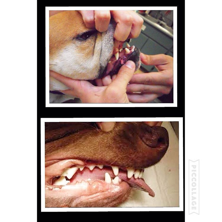
‘Base narrow’ canines (Linguoverted or ‘inverted’ canines) are a relatively common and painful problem in Australian dogs. The lower canines erupt more vertically or ‘straight’ than normal (instead of being tilted outwards), and strike the roof of the mouth. This causes pain whenever the dog chews or closes its mouth, and can result in deep punctures through the palatal tissues (sometimes the teeth even penetrate into the nasal cavity!). In our practice in Sydney, we see this most commonly in Staffordshire Bull Terriers and Labrador Retrievers.
Lance’ canines (Mesioverted, hard or ‘spear’ canines) occur when an upper canine erupts so it is pointing forward, like a tusk. This is seen most commonly in Shetland Sheepdogs, and can lead to lip trauma and displacement of the lower canine tooth (which cannot erupt to sit in its normal position in front of the upper canine).
Class II malocclusions (‘overshot’) arise when the lower jaw is relatively short compared with the upper jaw. This type of occlusion is NEVER considered normal and can result in significant and painful trauma to the upper gums, hard palate and teeth from the lower canines and incisors.
Class III malocclusions (‘undershot’, ‘prognathism’) occur when the lower jaw is relatively long compared with the upper jaw. The upper incisors may either meet the lower ones (level bite) or sit behind them (reverse scissor bite). While this is very common, and considered normal for some breeds, it can cause problems if the upper incisors are hitting the floor of the mouth or the lower teeth (similar problems to rostral crossbite). If the lower canines are striking the upper incisors, the accelerated dental wear often results in dead or broken teeth.
Class IV malocclusions (‘wry bite’) occur when there is a deviation of one or both jaws in any direction (up and down, side to side or front to back). These may be associated with mild to severe problems with chewing, damage to teeth and oral tissues, and chronic pain.
Normal development of the teeth and jaws is largely under genetic control, however environmental forces such as nutrition, trauma, dental interlock and other mechanical forces can also affect the final outcome.
As the interaction between these factors can be quite complex, it is recommended that you have your pup individually assessed – feel welcome to call me for advice.
Most malocclusions involving jaw length (skeletal) abnormalities are genetic in origin. We need to recognise this as it has enormous implications if you are planning to breed, as once a malocclusion is established in a line, it can be heartbreaking work to try and breed it back out.
The exact genes involved in jaw development are not yet well understood. We do know that the upper and lower jaws grow at different rates, at different times, and are under separate genetic control. In fact, the growth of one only affects the growth of the other if there is physical contact between them via the teeth. This contact is called ‘dental interlock’.
When the upper and lower teeth are locked against each other, the independent growth of either jaw is severely limited. This can occasionally work in the dog’s favour, for example if the lower jaw is slightly long compared with the upper jaw, the corner incisors may lock the lower canines in position behind them, limiting any further growth spurts of the lower jaw.
However, in many cases, dental interlock interferes with jaw development in a negative way. A classic example we see regularly in our practice is when a young puppy has a class II malocclusion (relatively short lower jaw) and the lower deciduous canines are locked behind the upper deciduous canines, or trapped in the tissues of the hard palate. In these cases, even if the lower jaw was genetically programmed to catch up to the upper jaw, it cannot physically do so.
Early removal of the lower canines (and often the lower incisors as well) to relieve this problem is strongly recommended. This procedure is called ‘interceptive orthodontics’ as we are ‘intercepting’ the developing problem before growth is completed and it is too late.
Extraction of these teeth will not stimulate jaw growth, but will allow it to occur if nature (ie genetic potential) permits. It also relieves the painful trauma caused by the teeth to the hard palate whenever the pup closes its mouth (and we all know how sharp those baby teeth are!!). More information on interceptive orthodontics can be found later in this book.
In some breeds, a genetic tendency for retained deciduous teeth can also contribute to the development of problems, such as anterior crossbite seen in several of the toy breeds.
It is crucial to remember that genetic malocclusions are not usually seen in all puppies in an affected litter as they are not dominant traits. Puppies can carry the genes contributing to genetic faults without showing any physical signs at all. If an affected puppy is noted, extreme caution should be exerted when planning future breeding from the parents and siblings, and neutering of the affected puppy is strongly recommended.
Although diet often gets the blame for development of malocclusions, the role of nutrition is actually much less significant than is often believed. Obviously gross dietary deficiencies will affect bone and tooth development, for example severe calcium deficiency can lead to ‘rubber jaw’. However, the vast majority of puppies are on balanced, complete diets and have adequate nutrient intake for normal bone and tooth development.
One myth I have heard repeated by several owners is that strict limitation of a puppy’s dietary intake can be used to correct an undershot jaw. This is simply NOT true. Limiting calories will NOT slow the growth of the lower jaw relative to the upper jaw (both jaws receive the same nutrient supply). Such a practice is not only ineffective, it can be detrimental for the puppy’s overall growth and development.
Trauma, infection and other mechanical forces may affect growth and development of the jaws and teeth. Developing tooth buds are highly sensitive to inflammation and infection, and malformed teeth may erupt into abnormal positions (or not erupt at all!). Damage to developing teeth can also occur if the jaw is fractured.
Retained or persistent deciduous (puppy) teeth can also cause malocclusions by forcing the erupting adult teeth into an abnormal position. As previously mentioned, this may be a genetic trait, but can also occur sporadically in any breed of dog.
A full bite assessment can help differentiate between malocclusions which are due to shifting of teeth alone, and those which have an underlying genetic basis. Contact me if you would like to arrange a bite assessment for your puppy
The basic rule is that every dog deserves a pain-free, functional mouth. If there is damage occurring to teeth, or oral tissues, we need to alleviate this, to allow the dog to live happily and healthily. If there is no functional problem and no trauma occurring, then treatment is simply not required.
Sometimes the hardest part is determining whether the problem is in fact causing pain. As we know, dogs are very adept at masking signs of oral pain, and will and will continue to eat despite real pain. Puppies, in particular, don’t know any better if they have had pain since their teeth first erupted very early in life.
Early assessment to determine whether intervention is required is critical in puppies with any signs of occlusal problems. Not only does this allow us to relieve their pain promptly, it can allow for easier correction of problems than if we wait until the permanent teeth have fully erupted and settled into place.
The overriding aim is always to give the dog a healthy, pain-free and functional mouth. Sometimes this will result in a ‘normal’ mouth, whereas in other cases, this might not be realistically achievable.
While some basic advantages and disadvantages of the different treatment options are outlined here, it is very important to seek specific advice for your individual dog, as no two mouths are exactly the same, and an individual bite assessment will help us determine the best course of action together. You can contact us anytime.
Malpositioned teeth may be moved into a more appropriate position using orthodontic appliances such as braces (yes, braces), wires, elastic (masel) chains or plates (similar to those used in humans!). In some cases, this may be a multi-step procedure which means repeated general anaesthetics.
Extraction of lower canine teeth – the roots of these teeth make up about 70% of the front of the jaw, and so there is a potential risk of jaw fracture associated with their removal. Some dogs also use these teeth to keep the tongue in position, so the tongue may hang out after extraction.
Extraction of teeth may severely limit an animal’s success in the show ring, especially in breeds where the correct number of teeth is emphasised in the breed standard.
Orthodontic movement of teeth is a complicated science, and, while some procedures appear quite straightforward, permanent damage to teeth and the surrounding structures can result from inappropriate procedures, poorly fitted appliances, or excessive pressures.
The outdated practice of using rubber bands to move the teeth is not recommended, as they slip down between the tooth and the gum, causing damage to the sensitive tissue here. The forces applied are also difficult to regulate, which can cause damage to the ligaments around the teeth, as well as the tooth roots. Much safer and more effective methods are now available.
Procedures to alter the shape of the teeth and make them fit better in the mouth can also be performed. This may vary from removal of small amounts of enamel (odontoplasty) to create space between teeth, right through to shortening the crown of a tooth to prevent it from causing trauma (crown reduction).
Crown reduction is commonly performed to treat base narrow canines, or class II malocclusions, where the lower canines are puncturing the hard palate. Part of the tooth is surgically amputated, a dressing inside the tooth to promote healing and the tooth is sealed with a white filling (just like the ones human dentists use). This procedure MUST be performed under controlled conditions as it exposes the highly sensitive pulp tissue. If performed incorrectly, the pulp will become infected and extremely painful for the rest of the dog’s life.
This pup has trauma in the roof of her mouth due to her left lower canine. A crown reduction procedure relieves the trauma while maintaining some functionality and avoiding extraction.
Even the less invasive odontoplasty (enamel shaping) can result in exposure of the sensitive dentine or pulp tissue if taken too far, and must be performed with extreme care to avoid permanent problems. Xrays are recommended prior to surgery so we can measure how far we can go before we get into the ‘danger zone’.
Although sometimes practised, clipping the tips of the teeth of puppies is NOT a humane procedure, and not only causes intense pain (imagine how it would feel if your own tooth was cut in half), but the resulting pulp infection can cause irreversible damage to the adult tooth buds which are developing underneath.
Extraction of teeth is sometimes performed, alone or in combination with other orthodontic treatments. This may be the preferred treatment in cases where:
While the dog may lose some function, this is far preferable to doing nothing (this condemns the dog to a life of pain). Indeed, unless released into the wild, dogs do well even if we need to extract major teeth (canines and carnassials), as they have the humans in their pack to do all the hunting and protecting for them.
This is the term we use when we remove deciduous teeth to alter the development of a malocclusion. The most common form of this is when we relieve dental interlock that is restricting normal jaw development. Such intervention does not make the jaw grow faster, but will allow it to develop to its genetic potential by removing the mechanical obstruction.
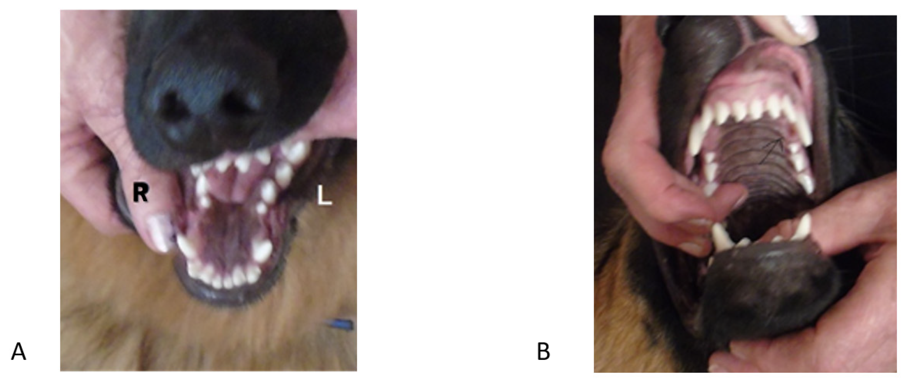
Extraction of deciduous lower canines and incisors in a puppy with an overbite releases the dental interlock and gives the lower jaw the time to ‘catch up’ (if genetically possible).
As jaw growth is rapid in the first few months of life, it is critical to have any issues assessed and addressed as soon as they are noticed, to give the most time for any potential corrective growth to occur before the adult teeth erupt and dental interlock potentially redevelops. Ideally treatment is performed from eight weeks of age.
Extraction of deciduous teeth is not necessarily as easy as many people imagine. These teeth are very thin-walled and fragile, with long narrow roots extending deep into the jaw. The developing adult tooth bud is sitting right near the root, and can be easily damaged. High detail intraoral (dental) xrays can help us locate these tooth buds, so we can reduce the risk of permanent trauma to them. Under no circumstances should these teeth be snapped or clipped off as this is not only inhumane, but likely to cause serious infection and ongoing problems below the surface.
Permanent enamel damage on adult teeth following extraction od deciduous teeth. The risk of this can be minimised by use of dental x-rays and extremely good surgical technique.
The aim of any veterinary procedure should always be to improve the welfare of the patient, so the invasiveness of any treatment needs to be weighed up against the likely benefits to the dog. Every animal deserves a functional, comfortable bite, but not necessarily a perfect one. Indeed, some malocclusions (particularly those involving skeletal abnormalities) can be difficult to correct entirely.
In addition to the welfare of the individual dog, both veterinarians and breeders need to consider the overall genetic health of the breed. Both the Australian National Kennel Club and (in New South Wales where our practice is situated) the Veterinary Practitioners’ Board stress that alteration of animals to conceal genetic defects for the purpose of improving their value for showing (and breeding) is not ethical.
For animals with malocclusions, very strong consideration needs to be paid to whether or not breeding from the affected animal is in the interests of improving the breed. If there is a genetic component, then neutering or selective breeding is recommended. As the vast majority of orthodontic abnormalities are not dominant in their inheritance (not all pups carrying the ‘bad’ genes will have visible problems), a ‘small’ issue seen sporadically can easily become widespread within a line.
This not only means many pups will have physical problems requiring correction for their own individual welfare, but breeding the problem out again can be extremely difficult.
The bottom line is that, while all dogs will have multiple treatment options available, and in some cases the occlusion can be corrected to the point of being ‘good for show’, advice should definitely be sought about the likelihood of a genetic component prior to embarking upon this, as the consequences for the breed can be devastating if such animals (or their close relatives) become popular sires or dams.
Sometimes a puppy may be missing one or more teeth. In the absence of trauma (which is usually apparent for other reasons!), there are a couple of things that may be going on.
Sometimes a tooth is congenitally missing, that is it has never developed. While dogs can physically cope well with missing teeth, in some breeds this is considered a serious fault, and will severely affect the chances of the dog being successful in the show ring.
Alternatively, a ‘missing’ tooth may be unerupted below the gumline. This can only be diagnosed using xrays. In some cases, the tooth may be trapped under a thickened layer of gum tissue, and surgery to relieve the obstruction (an operculectomy) may allow the tooth to erupt smoothly into the correct position if performed early enough.
Impacted lower canines trapped under thick gum tissue. They are also in a base narrow position. These teeth were able to erupt when tissue was surgically released (operculectomy).
Sometimes, the tooth will be in a favourable position but caught behind a small rim of jawbone – again early surgical intervention may be successful in relieving this obstruction. If the tooth is in an abnormal position or deformed, it may be unable to erupt even with timely surgery.
Impacted or embedded teeth should be removed if they are unable to erupt with assistance. If left in the jaw, a dentigerous cyst may form around the tooth. These can be very destructive as they expand and destroy the jawbone and surrounding teeth. Occasionally these cysts may also undergo malignant transformation (ie develop into cancer).
Firstly, if there are two teeth in one socket (deciduous and adult), the surrounding gum cannot form a proper seal between these teeth, leaving a leaky pathway for oral bacteria to spread straight down the roots of the teeth into the jawbone. Trapping of plaque, food and debris between the teeth also promotes accelerated periodontal disease. This not only causes discomfort and puts the adult tooth at risk of early loss, but allows infection to enter the bloodstream and affect the rest of the body.
If the deciduous tooth is still firmly in position as the adult tooth is erupting, it forces the adult tooth into an abnormal position which can cause a significant malocclusion. For example, the lower adult canines normally erupt on the inside of the deciduous teeth, so if they are forced to erupt alongside them, a painful base narrow malocclusion can result.
The upper adult canines normally erupt in front of the deciduous ones, so forcing them further forward can result in ‘lance’ canines. Finally, the upper adult incisors usually erupt behind their deciduous versions, so if these are retained a rostral crossbite may develop.
Retained upper baby canines force the adult canines to erupt in a more forward position. This can close the gap where the lower canine usually sits, forcing it into a traumatic position.
Puppies play rough, chew whatever they can get hold of, and have tiny teeth with very thin walls. Therefore fractures will sometimes occur. A common misconception is that broken deciduous teeth can be left until they fall out. Unfortunately this is NOT true. From the puppy’s point of view, broken teeth HURT, just as they do in children. Anyone who has had a bad toothache would agree that even a few weeks is a long time to wait for relief!
Broken teeth also become infected, with bacteria from the mouth gaining free passage through the exposed pulp chamber inside the tooth, deep into the underlying jawbone. This is not only painful, but can lead to irreversible damage to the developing adult tooth bud, which may range from defects in the enamel (discoloured patches on the tooth) through to arrested development and inability to erupt. The infection can also spread through the bloodstream to the rest of the body. Waiting for the teeth to fall out is NOT a good option!
We cannot rely on dogs to tell us when they have oral pain. It is up to us to be vigilant and watch for signs of developing problems. Train your pup to allow handling and examination of the mouth from an early age. We will be posting some videos of oral examination tips shortly, watch out in your email inbox for this. Things can change quickly – check their teeth and bite formation frequently as they grow.
Seek veterinary care as soon as a potential problem is noticed – you can call me on 1300 838 336 or email me anytime on support@sydneypetdentistry.com.au for advice or assistance.
Remember, early recognition and treatment is crucial if we want to keep your dog happy and healthy in and out of the show ring. The sooner we treat dental problems, the higher the chance of getting the best possible results with the least invasive treatment.

Occlusion is defined as the relationship between the teeth of the maxilla (upper jaw) and mandibles (lower jaw). When this relationship is abnormal a malocclusion results and is also called an abnormal bite or an overbite in dogs and cats.
The mouth is split into quadrants: left maxilla, right maxilla, left mandible and right mandible. Each quadrant of the mouth in both dogs and cats contains incisors (I), canines (C), premolars (PM) and molars (M).
In the normal, aligned mouth, the left and right side mirror each other. Dogs have a total of 42 adult teeth and cats have 30 adult teeth. The normal occlusion of a dog and cat mouth are similar. Below we"ll share how malocclusions can affect both canines and felines.
A malocclusion in that case, is either a tooth in an abnormal position and/or the misalignment of the maxilla and mandible with respect to one another."
A Class I malocclusion takes place when one or more teeth are in an abnormal position, but the maxilla and mandibles are in a normal relationship with each other. A Class I tooth may be pointing in the wrong direction or rotated.
Class III malocclusions are considered underbites in dogs and cats; the mandibles are longer in respect to their normal relationship to the maxilla. Class III malocclusions are commonly seen in brachycephalic dogs (boxers, pugs, boston terriers, etc).
Class IV malocclusions result from asymmetrical development of the maxilla or mandibles. The asymmetry of this malocclusion results in skeletal malformations leading to a side to side malalignment.
While cats do not get malocclusions nearly as frequently as dogs, they are not free from this problem. When present, felines malocclusions tend to be more severe and can cause more problems. There is a definitely a breed predisposition for cats with malocclusions. Persians and Himalayan cats tend to have a higher incidence of malocclusions, most frequently underbites.
Just as with dogs, cats with malocclusions should always be evaluated by a veterinarian and treated if their bite is traumatic and causing them pain. Malocclusions are frequently diagnosed in kittens. These abnormal bites are often painful and the sooner they are treated, the better the prognosis for gaining a pain-free and functional bite.
Malocclusions can result in an abnormal bite which can affect function and result in pain. Malocclusions predispose the patient to periodontal disease, endodontic (pulp) disease and oral trauma.
Our belief at Animal Dental Care and Oral Surgery is that all pets deserve a pain-free and functional bite. In most situations, the earlier a malocclusion is diagnosed the better the prognosis.
Class II and class III malocclusions are skeletal abnormalities resulting from abnormal development of the maxilla and mandible. It is rarely possible to restore the maxilla and mandibles to a normal relationship with each other, but it is always possible to permanently relieve pain that these malocclusions cause.
Although complete correction of certain teeth misalignments may not be possible, there is always something we can do to improve the functionality of the bite and make the patient comfortable. For best results, it is important to recognize a malocclusion as early as possible.
The upper incisor teeth slightly overlap the lower incisors. The lower canine tooth (fang) sits between the upper canine tooth and 3rd incisor. The premolar teeth do not touch each other and form zigzag-like pattern between the upper and lower premolars. The large upper 4th premolar tooth rests on the cheek side of the lower first molar. This tooth is also known as the upper carnassial tooth. The molars are not visible in this image.
Known as a canine overbite, the upper canine tooth is sitting in front of the lower canine tooth and is pointed forward. This is referred to as a mesioverted or “lance” canine tooth. Compare this image to the normal occlusion in figure 2.
Known as a canine underbite, the lower incisors are in front of the upper incisors and the lower canine tooth is resting against the back of the upper 3rd incisor. This bite is common in brachycephalic breeds, such as boxers and pugs.
Each side of the mandible developed at different rates resulting in a deviation to the left side. Note how the right lower canine is poking into the palate (roof) of the mouth. This is a significant source of pain for this patient.

These cookies are strictly necessary to provide you with services available through our websites. You cannot refuse these cookies without impacting how our websites function. You can block or delete them by changing your browser settings, as described under the heading "Managing cookies" in the Privacy and Cookies Policy.

Are you worried that you might have an underbite dog? For many dog breeds, underbites are relatively common. In some dogs they cause problems, in others they live quite happily never suffering as a result. This depends on the severity of the underbite, and some other factors too. Today I’m going to share the most popular underbite dog breeds. I’ll help you decide if your dog has an underbite, let and if it might cause an issue for them at some point.
When dogs have underbites, some people may find it cute or attractive. Unfortunately, there can be some trouble caused by canine malocclusion, the technical term for dog underbite conditions. Underbites are common in some breeds of dog. An underbite is when teeth are not aligned properly, causing the lower row to jut further out than the upper row.
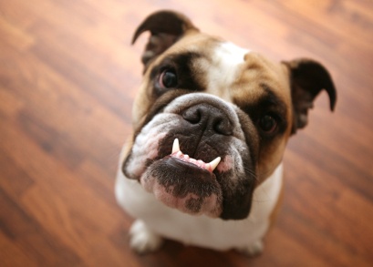
Common examples of this include the bulldog, who appears to have teeth protruding over his top lip. This will often result in the lower row of teeth being visible even when the dog’s mouth is closed. Underbites in dogs can range from very mild, requiring no action, to very severe, potentially requiring surgery.
In humans, identifying a clear underbite is quite simple, as we all have relatively similar jaw structures. There is a clear definition and appearance for what is “normal” when it comes to dental health. However, when it comes to canines, there is a less clear definition of “normal.”
There are a lot of variations from breed to breed and even litter to litter in some cases regarding the look of a dog’s teeth. Thus, identifying whether or not there will be dog underbite problems is not a question of what is “normal,” but rather what is comfortable and functional. If a dog has underbite visually, but they can functionally and comfortably chew food, there’s no need to worry.
On the other hand, if your dog has tooth-to-tooth or tooth-to-tissue contact that shouldn’t be there, it can cause pain and discomfort for your furry friend. True canine malocclusion must be diagnosed by a veterinarian or veterinary dental expert. In some cases, malocclusion can lead to other health issues. so it’s important to address it and discuss treatment options with a veterinarian.
It’s possible for any breed of dog to develop an underbite, but it’s much more common in certain breeds. Small dog breeds with underbites are most common, although some larger breeds like the boxer are also at risk. Small breeds such as the Boston terrier, Pekingese, French bulldog, English bulldog, King Charles Spaniel, Pug, Lhasa Apso and Shih Tzu are commonly observed developing underbites. These are the most common underbite dog breeds, but the condition is possible in most dogs. Also, keep in mind that mixed breed dogs with one or more parent breed from the list above will also be at risk for developing malocclusion.
A dog with an underbite may be at increased risk for various health problems. In cases of slight underbite due to a hereditary skeletal malocclusion, serious health issues are unlikely. As long as the dog is able to eat, drink and self-groom without issue, there is no cause for concern.
More serious cases of skeletal malocclusion can cause problems, however, as can dental malocclusions. Dogs with serious underbites can have difficulty chewing and swallowing food. This will usually be pretty easy to spot.
Misaligned teeth can also cause damage to gums and the soft tissues of the mouth. If unaddressed, this can result in discomfort for your dog and an increased risk of infection.
In severe cases, underbites can cause oronasal fistula, a condition in which a hole forms between the mouth and nose. This can cause severe pain, and in some cases even nasal disease and infection. Some warning signs to watch out for include signs of oral pain (such as the dog shying away when you pet its face), blood in saliva, abnormally bad breath, and difficulty eating or drinking.
Underbites in canines can fall into one of two categories: skeletal or dental. Dental malocclusion happens when a dog has a normal facial skeletal structure, but has one or more teeth that are abnormally positioned. Skeletal malocclusion occurs when the dog’s facial structure is abnormal. Resulting in the upper and lower rows of teeth not fitting together properly.
Both dental and skeletal malocclusion causes are at least somewhat genetic. Genetics can increase the likelihood of malocclusion, and these traits can be passed down between generations. Problems during gestation or early development can also lead to underbites, as can injuries and infections in puppies.
In some breeds, underbites are actually the result of intentional breeding practices. Some breeders may breed their pups specifically to engineer the type of jaw structure of a bulldog or a boxer. Like other questionable breeding practices, this crosses a line for some people. Underbites can cause discomfort and health problems for some dogs, so many believe that intentionally breeding underbite dog breeds is wrong.
If you are shopping around for puppies and have your eyes on one with an underbite, there are some things to consider before bringing them home. First and foremost, consider the potential health problems your pup may face later in life. Also, consider the fact that you might run into some larger-than-usual vet bills if serious problems do develop.
Although underbite dogs might seem endearing and arguably “cute,” your focus should be on the health and happiness of your pup—not just their cuteness factor. With all that said, keep in mind that slight underbites in puppies are not necessarily permanent.
This misalignment can sometimes self-correct as the dog develops. This is particularly true in some breeds with more pronounced muzzles, in which slight underbites are common. With that said, most small dogs that show symptoms as a young puppy will likely have a dog underbite for the rest of their lives. As a rule of thumb, the alignment of a dog’s teeth is typically permanent once it reaches about 10 months old—although this can vary from breed to breed.
Why does my dog have an underbite, and what should I do about it? If you have a dog with an underbite, it doesn’t matter much why it’s there. The important question is whether it’s causing your dog discomfort? This can often be difficult to tell. Most of the time the dog will have had the condition for his or her entire life, and won’t necessarily show signs of it bothering them.
In some cases, underbites don’t cause any irritation and are nothing to worry about. Or it could be causing your pup significant discomfort, even if he isn’t showing it. In any case, you should take your dog to the vet to have the underbite examined. A veterinarian will be able to tell the severity of the condition and check for signs of pain and infection.
From there, your vet will either recommend a treatment (see below) or let you know that no treatment is required. Even if you are given the all-clear by your vet, you should still keep an eye on your dog and watch for behavioral changes that may signal discomfort. Trouble eating, blood in the saliva, or signs of sensitivity around the mouth or nose should all trigger a return visit to your vet.
In many cases, no treatment will be recommended. Unless the underbite is causing discomfort or increasing the risk for disease, there is no need to correct it. If a vet determines that the dog underbite does require treatment, there are a few ways to go about it. Dog underbite correction options include removal of problem teeth, oral surgery or the use of an orthodontic appliance.
All of these treatments are pricey and invasive, so they should only be completed at the recommendation of your trusted veterinarian. In some cases, your vet will be able to do the work themselves but often they will refer you to an animal orthodontist or dental specialist.
This question, posed to me by a fellow passenger on my return flight from a veterinary conference, caused me to put my current task (subject-tagging images of dogs" and cats" mouths on my computer) on pause. I explained that in cats and dogs, the goal of orthodontic correction isn"t a pretty smile but pain-free, functional occlusion.
What happens when you peek into the mouth of a patient and note that one or more teeth are out of place? Hopefully you don"t quickly close the mouth, hoping that the pet owner didn"t spot the problem. (Out of sight, out of mind.) It"s much better to let your client know when something isn"t right in their pet"s mouth and explain what it will take to fix a poor or nonfunctional bite. But before you can recommend orthodontic care for your patients, you"ll need to embrace the concepts of malposition and malocclusion.
Occlusion refers to the relationship between the maxillary and mandibular teeth when they approach each other, as occurs during chewing or rest. Normal occlusion exists when the maxillary incisors just overlap the mandibular incisors (Figure 1A), the mandibular canines are equidistant from the maxillary third incisors and the maxillary canine teeth, and the premolar crown tips of the lower jaw point between the spaces of the upper jaw teeth in a saw-toothed fashion (Figure 1B). Flat-faced breeds, such as boxers, shih tzus, Boston terriers, Lhasa apsos and Persian cats, have abnormal bites that are recognized as normal for their breed in which the mandibular jaw protrudes in front of the maxillary jaw, altering the above tooth-to-tooth relationship (Figures 2A and 2B).
Malocclusion refers to abnormal tooth alignment. Skeletal malocclusion occurs when jaw anomalies result in abnormal jaw alignment that causes the teeth to be out of normal orientation. Dental malposition occurs when jaw alignment is normal but one or more teeth are out of normal orientation.
When dental malposition or skeletal malocclusion causes trauma to other teeth or oral soft tissues, the condition is termed poorly functional ornonfunctional and treatment is indicated. Therapy options include moving or removing the offending or offended tooth or teeth, or surgically creating additional space for the malpositioned tooth to occupy without causing trauma.
Mandibular distoclusion (also called overbite, overjet, overshot, class 2, and mandibular brachygnathism) occurs when the lower jaw is shorter that the upper and there"s a space between the upper and lower incisors when the mouth is closed. The upper premolars will be displaced rostrally (toward the nose) compared with the lower premolars. Mandibular distoclusion is never normal in any breed (Figures 3A and 3B).
Figure 3B. A dog"s mandibular distocluson.Mandibular mesioclusion (also called underbite, undershot, reverse scissor bite, prognathism, and class 3) occurs when the lower teeth protrude in front of the upper teeth. If the upper and lower incisor teeth meet each other edge to edge, the occlusion is an even or a level bite (Figure 4).
Figure 4. Mandibular mesioclusion in a dog.Maxillary mandibular asymmetry (also called wry bite, especially by breeders) is a skeletal malocclusion in which one side of the jaw grows differently from the other side (Figures 5A and 5B).
Rostral cross bite occurs when the canine and premolar teeth on both sides of the mouth are normally aligned but one or more of the lower incisors are positioned in front of the upper incisors (Figure 6).
Figure 6. Rostral cross bite.Mesioverted mandibular canines (also called lingually displaced canines or base narrow canines) occur when the lower canine teeth protrude inward, impinging on or penetrating the maxillary gingiva (Figure 7). Often this condition is due to retained deciduous teeth. The resulting trauma can be alleviated through tooth movement, crown reduction and restoration, or extraction.
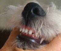
You can use this royalty-free photo "Small dog with undershot jaw posing indoors" for personal and commercial purposes according to the Standard or Extended License. The Standard License covers most use cases, including advertising, UI designs, and product packaging, and allows up to 500,000 print copies. The Extended License permits all use cases under the Standard License with unlimited print rights and allows you to use the downloaded stock images for merchandise, product resale, or free distribution.

A dog"s bottom jaw will continue growing until the age of 10 - 12 months. Until this time a slight undershot will often correct itself naturally with ageing. A severe overshot is unlikely to correct itself but is not a significant health issue for the dog. Overshot dog"s can still lead healthy lives as happy and loving pets regardless of this problem. Dog"s with an overbite should not be shown or bred from.UNDERSHOT BITE:also referred to as an underbite. This when the bottom jaw is longer than the top jaw and the incisors on the bottom jaw protrude past those of the top jaw. This type of bite is actually correct for breeds such as the bulldog. An underbite generally does not adversly effect the dog in anyway. These dogs still make loving and healthy pets but should not be shown or bred from.LEVEL BITE:also called the pinscer bite. This is where both jaws are the same length and the incisors meet edge to edge. This bite does not affect the dog adversely in any way but can cause premature wearing of the incisor teeth. Often in a slightly older dog the bite can go from scissor to level with ageing.WRY BITE:This is where the bottom jaw is twisted and the incisors do not meet in a correct straight scissor bite. This type of bite is not very common and generally the dog suffers no ill effects. A dog with a wry mouth should not be shown or bred from.
In most cases and overshot, undershot, level or wry bite is not a serious condition and should not discourage someone from purchasing such a pup unless their intentions are specifically to show or breed. In the wild a dog with an incorrect bite could have great difficulty hunting and killing prey. For the purposes of a pet an incorrect mouth is not a serious concern as modern prepared dog foods are in palatable sized portions. An incorrect bite would however exclude a dog from a working career where the dog was required to use it"s mouth, for example, herding or police work.BITE GENETICSThe inheritance mode of a dog"s bite is largely unknown and litters may present with confusing outcomes. However, because an incorrect bite can be determined by 12 months of age, a dog with such a fault can be excluded from a breeding programme.
Incorrect bites vary greatly with respect to severity and occurrence. Some breeds and lines within breeds appear to have a higher incidence of bite faults than others. It has also been found that dogs with undershot bites often result from parents with correct scissor bites. This would indicate that the fault is recessive but no conclusive proof is available.
Dogs who produce offspring with bite faults should not necessarily be exclude from a breeding programme because of this reason alone. There is a saying, "Don"t throw out the baby with the bathwater." Which basically means that a good example of the breed both in conformation and temperament should not be excluded solely because it has produced offspring with an incorrect bite. Other offspring from this dog may be perfectly correct in mouth. To exclude such a dog would mean eliminating valuable genetic material from the population unnecessarily. To breed from this dog would however mean that the fault can not be eliminated completely from future generations and as such the offspring would need to be checked for this fault. Often a recessive fault can go unseen for several generations before it makes an appearance again.
It has also been suggested that the bite might not be entirely governed by genetics and that the size of the actual incisors can play a role in the bite. In our own breeding programme we have observed that dogs with larger incisors are less likely to have an even or overshot bite as an older dog.
Because the bottom jaw continues to grow until the dog reaches 12 months is has been observed that a puppy with a slight overshot bite has corrected. Hence it is sometimes worth retaining an otherwise promising puppy that may have a very slight gap in the jaws at a young age. Some people say that a matchstick gap is ideal. Puppies with smaller incisors and no gap can actually go even or undershot. Some breeders also believe that a slight overshot can be corrected by administering the puppy extra calcium supplements at a young age while the jaw is still growing.
A bite can stay the same throughout puppyhood or change greatly as the dog grows during the first year. There is no hard and fast rule. We have observed a particular puppy go from being overshot to scissor to even to undershot in the space of several months while it"s littermates held perfect scissor bites the whole time.MISSING TEETH (incomplete dentition)Another concern from a breeding perspective is dogs that have missing teeth. For most working and herding breeds the standard requires full dentition (42 teeth).
Once again the genetic mode of inheritance for missing teeth is unknown. Parents with full dentition can produce offspring with one or more missing teeth suggesting inheritance is recessive. This appears to be compounded by the fact that x-rays have shown that some dogs who were thought to have a missing tooth, actually possessed the tooth in question but it was below the gum-line and was therefore not visible.
Some breeders are overly pedantic about a dog having one missing tooth and will eliminate such a dog (and even sometimes it"s parents) from their breeding programme. This action is unwarranted as the genetic material in an otherwise excellent specimen could be lost forever. This decision can be yet another example of "Throwing out the baby with the bathwater." Particularly when the mode if inheritance is not understood.
Although missing teeth are certainly not desirable in a show or breeding dog, there are very few standards that actually describe this as a serious or disqualifying fault (one of which is the German Shepherd Dog). The ANKC standards for the Australian breeds do not state that full dentition is required therefore although not desirable, a dog should not be penalised for having a missing tooth.MOUTH HEALTHA puppy"s teeth and bite should be regularly checked whilst they are growing. This is to ensure that the bite is correct but also to make sure that the deciduous teeth fall out correctly as the permanent teeth grow in. It is possible for a puppy to retain baby teeth particularly the canine teeth (this is quite common in smaller breeds). These teeth will need to be pulled or removed if they don"t fall out naturally. Sometimes the tooth is already loosened by the permanent tooth and can just be wiggled and will come free. It the tooth is still deep rooted and not loose it may need to be removed by a veterinarian.
When puppies are teething their gums are often sore and swollen. It is of value to supply the puppy with something suitable and safe to chew which will both help relieve the discomfort and loosen the deciduous teeth. Brisket bones and rawhide chews are ideal.
Overall gum and tooth health requires the owner to periodically check the teeth for build up of tartar or any infections. A dog"s teeth need to be checked just like humans do. It is also worth mentioning that a diet of only soft foods is not good for tooth health.

teeth so those tiny teeth need to be pulled at the appropriate time. Experience has taught me that few vets are knowledgeable about canine dentistry.
Basically, toy breeds" lower jaw grows faster than the upper jaw. This causes the bite to go undershot (the lower teeth protrude beyond the upper teeth – a reverse scissor bite or worse). A scissor bite is a “man-made” bite and the even bite is a natural bite. So according to one of the six board certified vets in canine dentistry, we have created our own problems with bites/teeth.
If a bite on a 12 week old puppy seems to be going off – (1) undershot; (2) overshot; or wry mouth, the appropriate teeth need to be removed – either upper and lower jaw or the teeth in the jaw that are binding. If the bite is undershot, then remove the upper jaw teeth; if the bite is overshot, remove the lower jaw teeth; if the bite is wry*, the canines should be removed because they are locking in place the way the teeth are coming in.
Extract the appropriate teeth of a questionable bite at 10 to 12 weeks of age.��Remove the baby teeth as soon as you see a bite starting to go off and certainly all baby teeth should be out by 6 months of age. The canine teeth don’t normally come out till the dog is 9 months of age but if the bite looks like it is off, the proper jaw’s canines should be removed.
If the dog is a valuable show dog, you run a risk of a messed up bite if you do nothing or your vet doesn’t know what to do or advises you to just watch the bite. It is safer to pull the teeth at the appropriate times.
A vet using isoflorine is best and no other medication, and one that can keep small kittens alive during surgery is what you need to find to do your dental work on toy breeds. If a vet says he/she is an expert on canine dentistry, ask to see board certification for having received education in this specific field and check to see if that expertise also includes experience with toy breeds.
*If the bite is a wry mouth, that is considered a “severe” problem and all the baby teeth on that side need to be pulled – even the back teeth. In slight under or over bites, remove only the front teeth and certainly remove the baby canines if they are binding any incorrect bite because they don’t normally come out on their own (if they come out at all) till 9 months of age. If the over or under bite is severe (quite a wide gap), then remove all the teeth in the appropriate jaw.
**If the bite is almost horizontal or “parrot bite,” you may have a depletion (leeching) of the calcium from the bones (particularly noticeable in the extremities and teeth) going on in your puppy. Feeding a balance (I stress BALANCE) of calcium/phosphorus and getting the baby teeth out (all of them because this is a severe condition) will correct that bite if the dog had a correct bite at 8 weeks (young puppy). I give OsteoForm from ages 3 - 4 months to 9 months or longer and it is especially necessary in puppies who have big growth spurts.
I pass this information on as what I have learned the hard way and it has taken me years to learn it through trial and error (way too many errors) so that you will not have to learn the hard way as I have and you will save many wonderful puppies that would have been lost as stars in the show ring.
If we know the dental formulas and eruption times, it will help us identify abnormal pathology including missing teeth, delayed eruption, persistent/retained teeth, and supernumerary teeth. Neither the first premolar nor any of the molars have deciduous teeth (they are nonsuccessional teeth). When differentiating between deciduous or permanent dentition, the deciduous teeth can often be identified based on the tooth shape and smaller size. It is important to remember that the deciduous third and fourth premolars will look like the adult fourth premolar and first molar in shape.
Common pathology associated with deciduous teeth include persistent or retained teeth. These two terms are often used interchangeably, however, this is not appropriate as each term is specific. Persistent deciduous teeth is the term that should be used for a deciduous tooth which remains present when the adult tooth is erupted. It persists, despite the presence of the adult tooth. The deciduous tooth in this case should be extracted. No two teeth should occupy the same space. The presence of the deciduous tooth can lead to malocclusion and will predispose the adult tooth to periodontal disease. The term retained is better suited for dental structure that is present underneath the gums (i.e. retained tooth root) or can sometimes be used when describing a deciduous tooth which is present due to the lack of an adult counterpart. I most commonly see this with second premolars in dogs. I let owners know that a dental radiograph should be performed to ensure there is no adult tooth. The deciduous tooth can remain if there is no indication of pathology associated with it. Owners are warned that the tooth roots may resorb and there is the potential the tooth can fracture – both situations requiring extraction in the future.
With adult patients we have more treatment options for a fractured tooth with pulp exposure (complicated fracture, endodontic disease) including extraction, root canal therapy, or vital pulpotomy given the right circumstances. If a deciduous tooth is fractured, treatment recommendation should be for extraction. Not only is the fractured tooth a source of pain and infection, but the inflammation associated with endodontic disease can affect the developing adult tooth bud. The apex of the deciduous tooth root sits in close proximity to the permanent tooth bud and changes in the local environment can lead to disruption of the ameloblasts forming enamel on the adult tooth.
Most times patients presenting for malocclusion will be pediatric or young juvenile when this issue is noticed. When assessing occlusion, keeping in mind the standards of normal occlusion are most important. Normal or ideal occlusion (canine) consists of interdigitation of the upper and lower teeth, maxillary incisor teeth are slightly rostral to the mandibular incisor teeth with the mandibular incisor teeth contacting the cingulum of the maxillary incisors, the mandibular canine teeth should be slightly inclined labially and sit within the interdental space between the maxillary third incisor and canine tooth, the crown cusps of the mandibular premolars should be lingual to the maxillary premolar teeth, the premolars should have a pinking shears relationship with the mandibular first premolar rostral to the maxillary premolars, the mesial cusp tip of the maxillary fourth premolar should be lateral to the space between the mandibular fourth premolar and first molar. Normal occlusion in cats follows the same basic guidelines with minor adjustments.
Malocclusions can be a result of abnormal tooth positioning or due to abnormal jaw length relationship. Many animals with malocclusion may have more than one deviation from normal and thus multiple concurrent malocclusions (i.e. mandibular distoclusion with linguoverted mandibular canine teeth). Class 1 malocclusion consists of a normal jaw length relationship with malpositioning of one or more teeth. Generally these malocclusions are described by the physical direction that the tooth is angled or deviated (i.e. mesioverted). Another version of class one malocclusion is a crossbite, where the mandibular teeth are more buccal or labial to the opposing maxillary tooth. Crossbites can be described as rostral crossbite – referring to the incisor teeth, or caudal crossbite – when the malocclusion is associated with the premolars or molars. Other classes of malocclusion deal with skeletal malocclusion.
Class 2 malocclusion, or mandibular distoclusion, is when the mandible occludes caudal to the normal position relative to the maxilla. Colloquial terms for this malocclusion include parrot mouth, overbite, overshot jaw. On the other hand, class 3 malocclusion, mandibular mesioclusion, the mandible will be rostral to the maxillary arch. In some breeds this occlusion is normal for the breed (i.e. brachycephalics). Other terms often used are underbite or monkey mouth. Class 4 malocclusions involve asymmetry and can be in a rostro-caudal, side-to-side, dorso-ventral direction, or a combination of these. We try to steer away from the term wry bite, as this is non-specific and does not accurately provide an image of a patient’s malocclusion. An example of class 4 malocclusion in a rostrocaudal direction would be when either the right or left side of the face has mandibular mesioclusion or distoclusion. A side-to-side malocclusion would describe when the midline alignment of the maxilla and mandible is shifted. And finally, a dorsoventral malocclusion would correspond with an open bite in which there is abnormal vertical space between the maxilla and mandible when the mouth is in a closed position.
Treating malocclusion cases can be more of an art than a science, and frequently we are dealing with evolving circumstances. Expectations of malocclusion treatment should be relayed to the owner and procedures should be performed to provide pets with a comfortable bite rather than cosmetic outcome. Options for treatment include interceptive orthodontics (extractions), corrective orthodontics, or alteration to a tooth shape/structure (crown reduction with vital pulpotomy).
Our role as veterinarians in helping pets have a mouth free of discomfort and disease should begin when our patients are very young. Issues with deciduous dentition should not be overlooked due to the short timespan that these teeth are present, since these issues may contribute to problems when the patient gets older. The stage when adult teeth are erupting is a crucial time to ensure that teeth are accounted for, structurally normal, and positioned appropriately.

This website is using a security service to protect itself from online attacks. The action you just performed triggered the security solution. There are several actions that could trigger this block including submitting a certain word or phrase, a SQL command or malformed data.
Every dog breed encounters health problems and Weimaraners are no exception. The below is a partial listing of some health issues that may affect Weimaraners. Note that only some, but not all of these are hereditary, genetic, and congenital conditions.
If you are searching for a Weimaraner puppy, please discuss these health anomalies with your breeder. Ask them if they’ve encountered any of these health woes, and how they’ll help new puppy owners address these issues if their puppy has or develops any of these conditions:
Cervical Spondylomyelopathy (CSM), or Wobbler Syndrome. This is a disease of the cervical spine (at the neck) that is commonly seen in large and giant-breed dogs. CSM is characterized by compression of the spinal cord and/or nerve roots, which leads to neurological signs and/or neck pain. The term wobbler syndrome is used to describe the characteristic wobbly gait (walk) that affected dogs have. Symptoms include a strange, wobbly gait, neck pain and stiffness, weakness, possible short-strided walking, spastic with a floating appearance or very weak in the front limbs, inability to walk, partial or complete paralysis, possible muscle loss near the shoulders, worn or scuffed toenails from uneven walking, increased extension of all four limbs, and difficulty getting up from a lying position. Causes: nutrition in some cases (excess protein, calcium and calories in the case of Great Danes), or fast-growth. Wobbler syndrome is diagnosed via visualization. X-rays, myelographs, computed tomography (CT) and magnetic resonance imaging (MRI) will allow your doctor to view the spine and vertebrae. X-rays should be used mainly to rule out bony disorders while myelographs, CT and MRI are used to visualize the compression of the spinal cord. Diseases that will need to be ruled out though a differential diagnosis include diskospondylitis, neoplasia, and inflammatory spinal cord diseases. The results of the cerebral spinal fluid (CSF) analysis should pinpoint the origin of the symptoms.
Congenital Diaphragmatic Hernia (or Congenital Diaphragmatic Rupture) is a tear in the muscle that separates the abdomen and the chest. In Weimaraners, we see this as a peritone


 8613371530291